©2007 Baishideng Publishing Group Co.
World J Gastroenterol. Feb 21, 2007; 13(7): 1108-1111
Published online Feb 21, 2007. doi: 10.3748/wjg.v13.i7.1108
Published online Feb 21, 2007. doi: 10.3748/wjg.v13.i7.1108
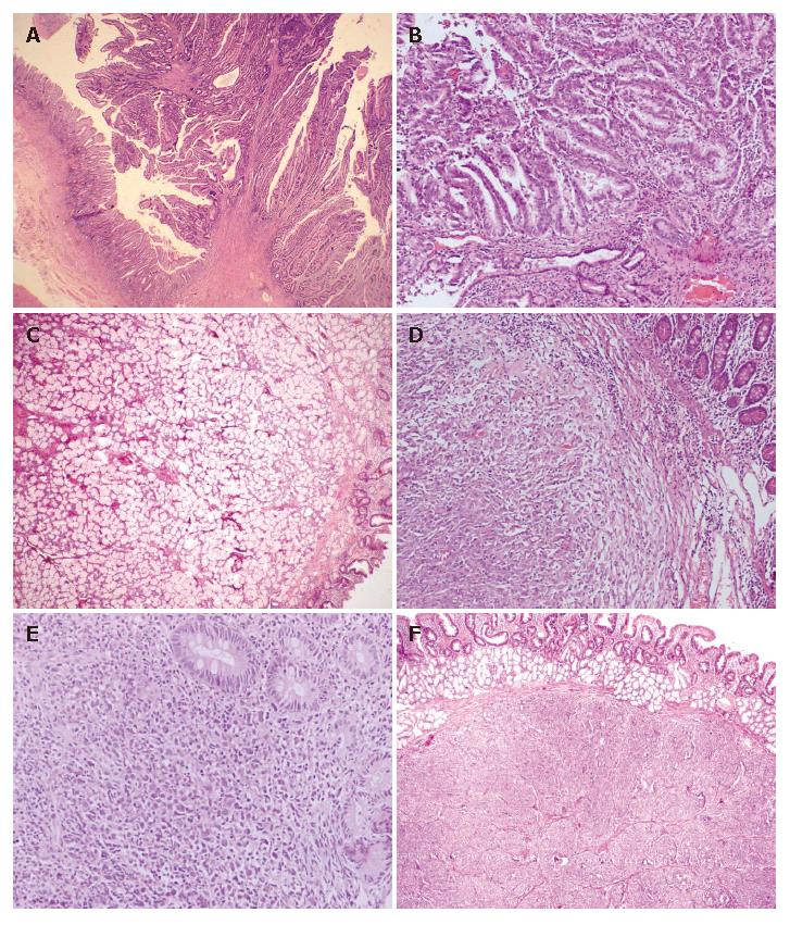
Figure 1 Photomicrograph showing tubulovillous adenoma of the duodenum (A) (HE × 100), adenocarcinoma arising in tubulovillous adenoma of the duodenum (B) (HE × 200), Brunner glands separated by fibrovascular septa in Brunner gland adenoma of the duodenum (C) (HE × 40), spindle cell gastrointestinal stromal tumour in submucosa of the duodenum (D) (HE × 100), non-Hodgkin’s lymphoma showing diffuse monotonus population of large cells infiltrating duodenal glands (E) (HE × 100), and circumscribed tumour nodule with tumour cells arranged in organoid pattern in carcinoid of the duodenum (F) (HE × 40).
- Citation: Bal A, Joshi K, Vaiphei K, Wig J. Primary duodenal neoplasms: A retrospective clinico-pathological analysis. World J Gastroenterol 2007; 13(7): 1108-1111
- URL: https://www.wjgnet.com/1007-9327/full/v13/i7/1108.htm
- DOI: https://dx.doi.org/10.3748/wjg.v13.i7.1108


