©2007 Baishideng Publishing Group Co.
World J Gastroenterol. Feb 14, 2007; 13(6): 960-963
Published online Feb 14, 2007. doi: 10.3748/wjg.v13.i6.960
Published online Feb 14, 2007. doi: 10.3748/wjg.v13.i6.960
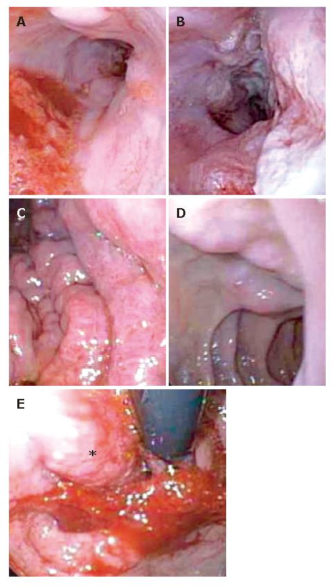
Figure 1 Esophagogastroduodenoscopy demonstratring grade 1 esophageal varices (A), tumor invasion of distal third of the esophagus (B), moderate portal hypertensive gastropathy (C), duodenal varices (D), retroflexed view of the nodular tumor in the gastric fundus (E).
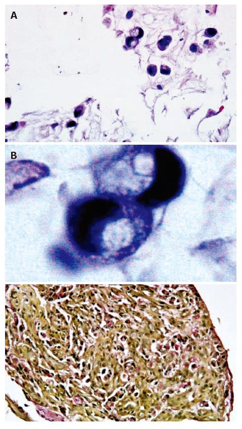
Figure 2 Histology and immunohistochemistry of biopsies from gastric: Light microscopy with hematoxylin and eosin staining of tumor tissue at 40X and 100X with oil magnifications demonstrating moderately-sized cells with abundant vacuolated cytoplasm (A) and occasional signet ring cells (B), and mucicarmine stain demonstrating intracytoplasmic positive staining (C).
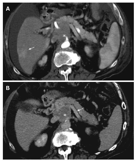
Figure 3 Contiguous helical CT scan of the abdomen at 5 mm collimation with intravenous contrast captured in arterial phase (A) and portal venous phase (B) at the level of the pancreas.
A 5 cm soft tissue mass (star) was seen with severe attenuation of the splenic (B, arrow), main portal and superior mesenteric veins. An area of increased attenuation within the segment VI of the liver was visualized in arterial phase (A, arrow) concerning for metastasis.
- Citation: Ghosh P, Miyai K, Chojkier M. Gastric adenocarcinoma inducing portal hypertension: A rare presentation. World J Gastroenterol 2007; 13(6): 960-963
- URL: https://www.wjgnet.com/1007-9327/full/v13/i6/960.htm
- DOI: https://dx.doi.org/10.3748/wjg.v13.i6.960


