©2007 Baishideng Publishing Group Co.
World J Gastroenterol. Feb 7, 2007; 13(5): 813-815
Published online Feb 7, 2007. doi: 10.3748/wjg.v13.i5.813
Published online Feb 7, 2007. doi: 10.3748/wjg.v13.i5.813
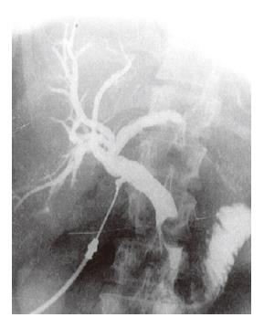
Figure 1 Showing filling defect within the distal common bile duct causing almost complete obstruction.
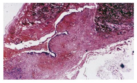
Figure 2 A pigmented malignant tumour evidently infiltrates extrahepatic bile duct.
Later histochemical and immunohistochemical analyses showed melanoma cells (HE, × 13).
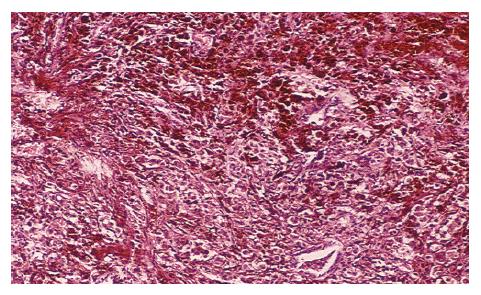
Figure 3 Higher magnification of the same tumour field showed insular and trabecular-to-solid histological organization of melanoma cells.
Most of the cells were hyperpigmented with massive intracytoplasmic melanosome granules (HE, × 64).
- Citation: Colovic RB, Grubor NM, Jovanovic MD, Micev MT, Colovic NR. Metastatic melanoma to the common bile duct causing obstructive jaundice: A case report. World J Gastroenterol 2007; 13(5): 813-815
- URL: https://www.wjgnet.com/1007-9327/full/v13/i5/813.htm
- DOI: https://dx.doi.org/10.3748/wjg.v13.i5.813


