Copyright
©2007 Baishideng Publishing Group Co.
World J Gastroenterol. Dec 28, 2007; 13(48): 6593-6597
Published online Dec 28, 2007. doi: 10.3748/wjg.v13.i48.6593
Published online Dec 28, 2007. doi: 10.3748/wjg.v13.i48.6593
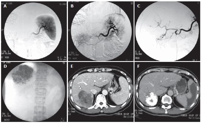
Figure 1 PSE treatment for a 68-year-old male case of HCC with splenomegaly and thrombocytopenia.
A: Splenic arteriography before PSE showing the whole splenic parnchymal image; B: Splenic arteriography after PSE showing the residual splenic parnchymal image, part of the peripheral splenic parenchyma was ablated, and the extent of embolization was roughly estimated of approximately 60%; C: Celiac arteriography before TACE showing the tumor blood-supply image; D: TACE is terminated when the tumor is filled with emulsifier; E: Transverse CT image revealing splenomegaly at 1 wk before PSE/TACE; F: Transverse CT image revealing the infarction of peripheral splenic parenchyma at 2 wk after PSE. The extent of embolization was 62% calculated by CT volume analysis software.
- Citation: Huang JH, Gao F, Gu YK, Li WQ, Lu LW. Combined treatment of hepatocellular carcinoma with partial splenic embolization and transcatheter hepatic arterial chemoembolization. World J Gastroenterol 2007; 13(48): 6593-6597
- URL: https://www.wjgnet.com/1007-9327/full/v13/i48/6593.htm
- DOI: https://dx.doi.org/10.3748/wjg.v13.i48.6593


