Copyright
©2007 Baishideng Publishing Group Inc.
World J Gastroenterol. Nov 14, 2007; 13(42): 5659-5661
Published online Nov 14, 2007. doi: 10.3748/wjg.v13.i42.5659
Published online Nov 14, 2007. doi: 10.3748/wjg.v13.i42.5659
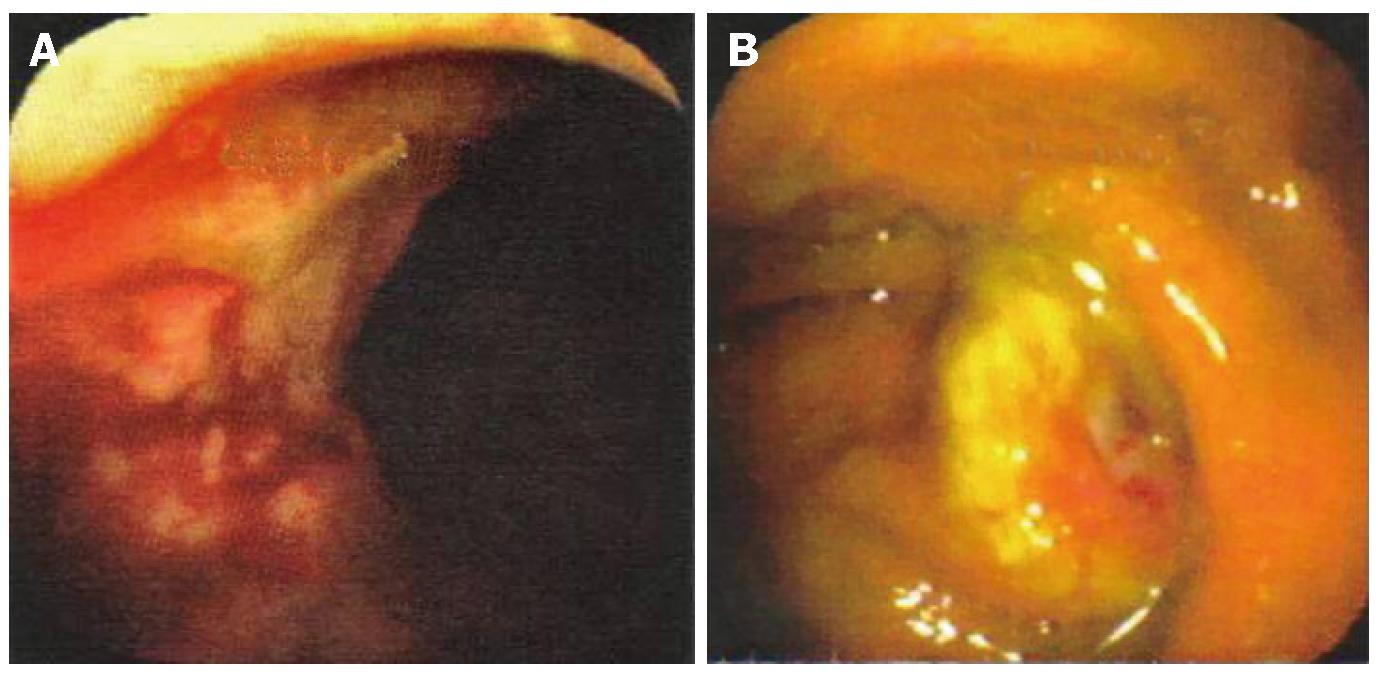
Figure 1 Endoscopy demonstrating ulcerated rectal (A) and cecal (B) lesions suggestive of carcinoma.
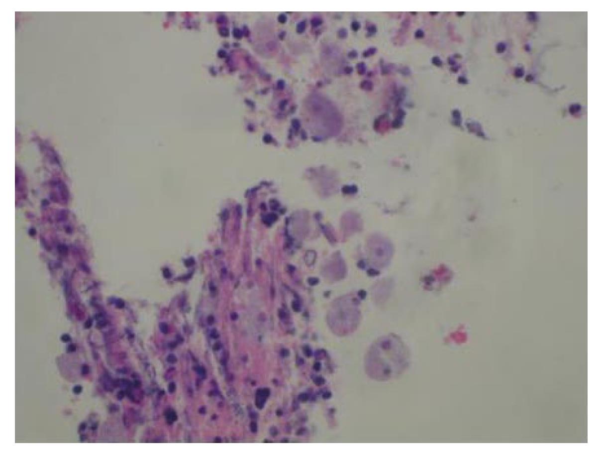
Figure 2 High-powered magnification of a hematoxylin-eosin preparation of a colonic biopsy that demonstrates trophozoites of E.
histolytica.
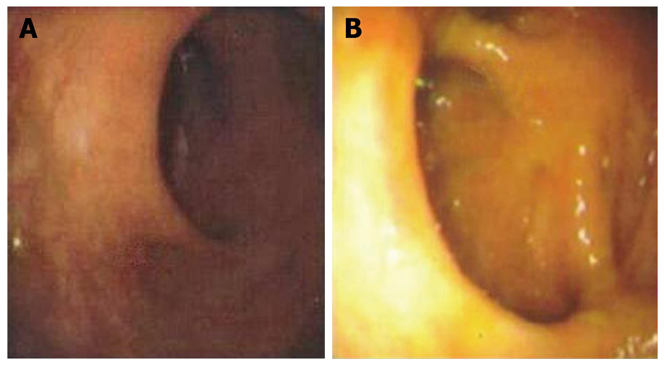
Figure 3 Follow-up colonoscopy subsequent to a completed course of antibiotic therapy, which demonstrates complete resolution of rectal and cecal lesions, with normal appearing colonic mucosa.
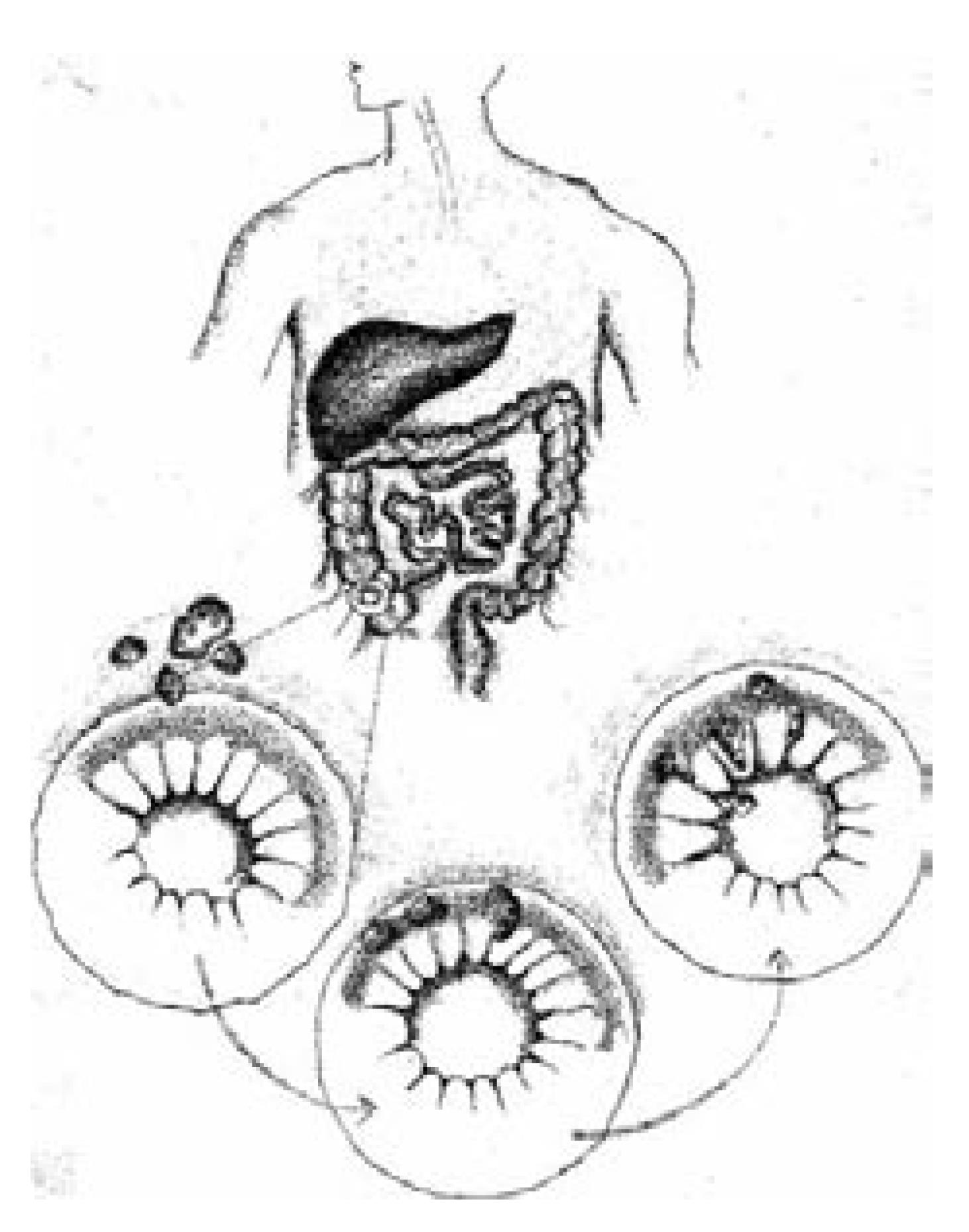
Figure 4 Pathogenesis of ameboma formation: infection is initiated by ingestion of fecally contaminated food or water containing E.
histolytica cysts. Excystation occurs in the bowel lumen in which motile and potentially invasive trophozoites are formed. Invasion of the mucosa and submucosa may lead to colitis. Ameboma forms when severe inflammatory reaction occurs, with formation of granulation tissue that leads to a pseudotumor appearance.
- Citation: Hardin RE, Ferzli GS, Zenilman ME, Gadangi PK, Bowne WB. Invasive amebiasis and ameboma formation presenting as a rectal mass: An uncommon case of malignant masquerade at a western medical center. World J Gastroenterol 2007; 13(42): 5659-5661
- URL: https://www.wjgnet.com/1007-9327/full/v13/i42/5659.htm
- DOI: https://dx.doi.org/10.3748/wjg.v13.i42.5659


