Copyright
©2007 Baishideng Publishing Group Co.
World J Gastroenterol. Nov 7, 2007; 13(41): 5521-5524
Published online Nov 7, 2007. doi: 10.3748/wjg.v13.i41.5521
Published online Nov 7, 2007. doi: 10.3748/wjg.v13.i41.5521
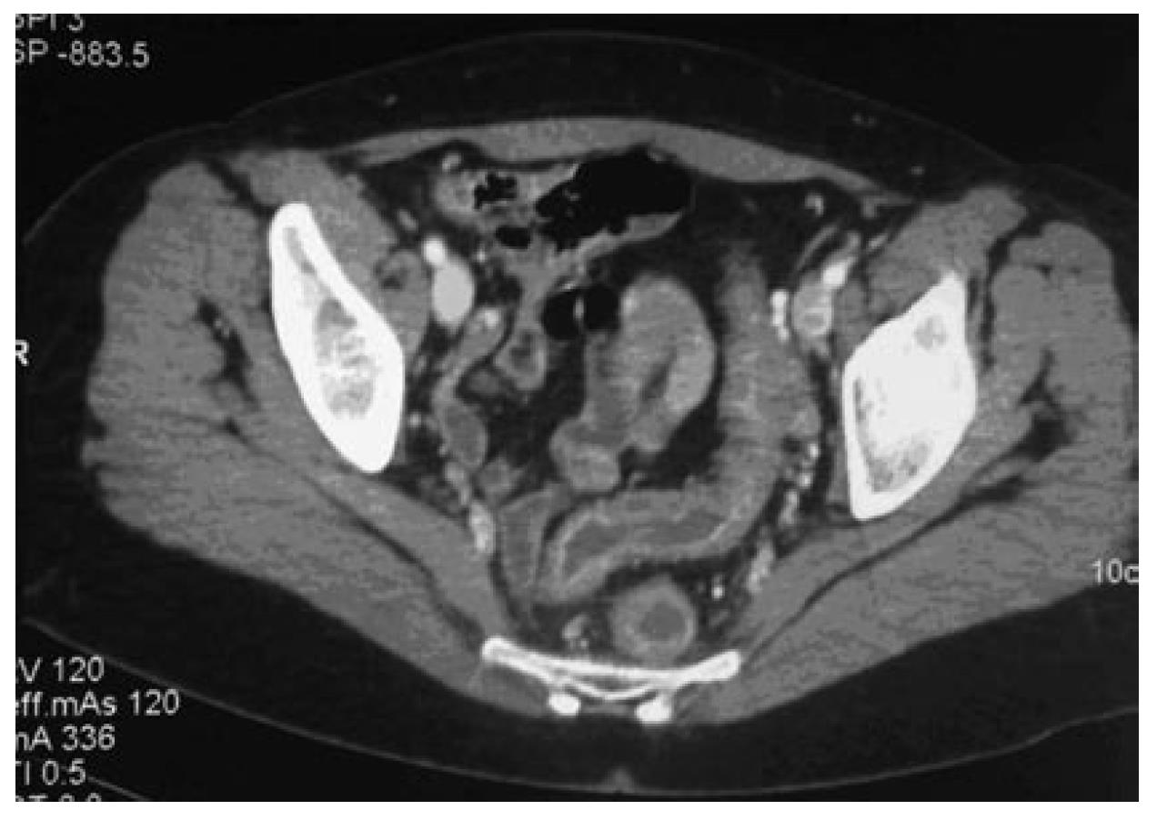
Figure 1 Contrast-enhanced CT scan of the abdomen showing a diffuse infiltration around the rectum and the sigmoid colon, and thickening of their walls.
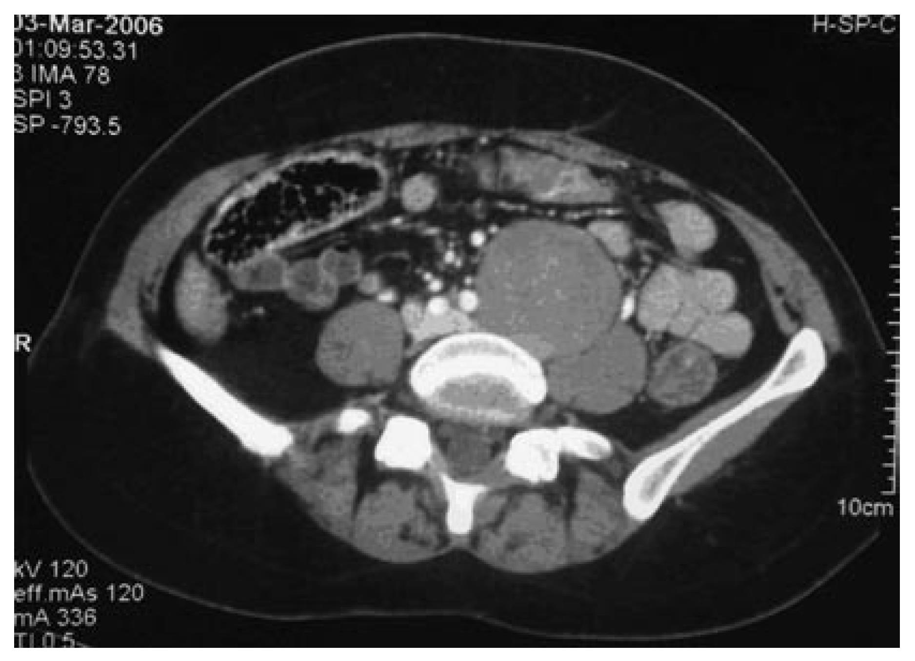
Figure 2 Well-demarcated, homogeneous mass measuring 60×50 mm in close proximity to the left iliac artery, lumbar vertebrae and psoas muscle, on contrast-enhanced CT scanning.
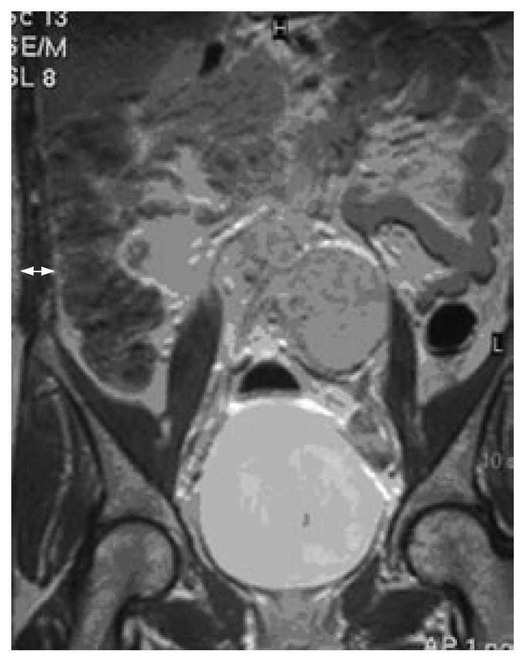
Figure 3 Coronal T1-weighted MR image using gadolinium, showing a solid mass with the same features as seen with CT scanning.
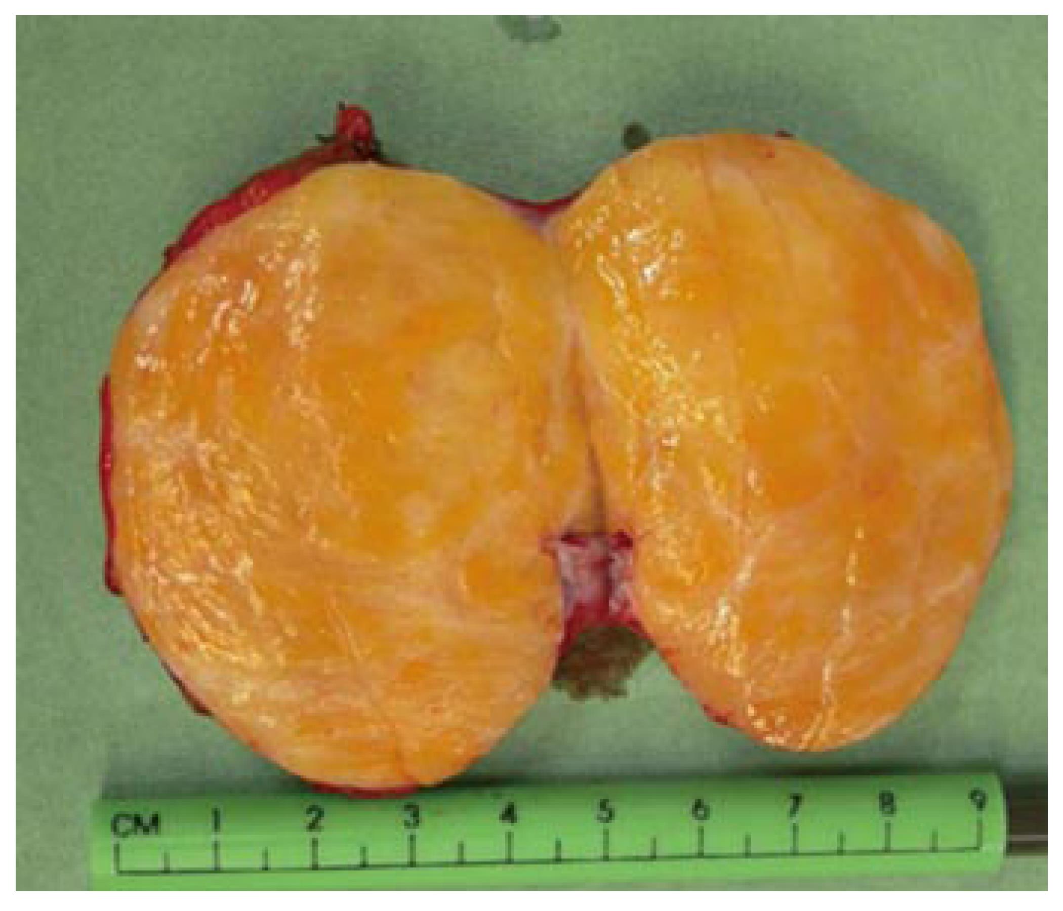
Figure 4 Perioperative examination of the mass revealed a solid, greyish, ovoid tumor with a smooth capsule and a homogeneous yellow core.
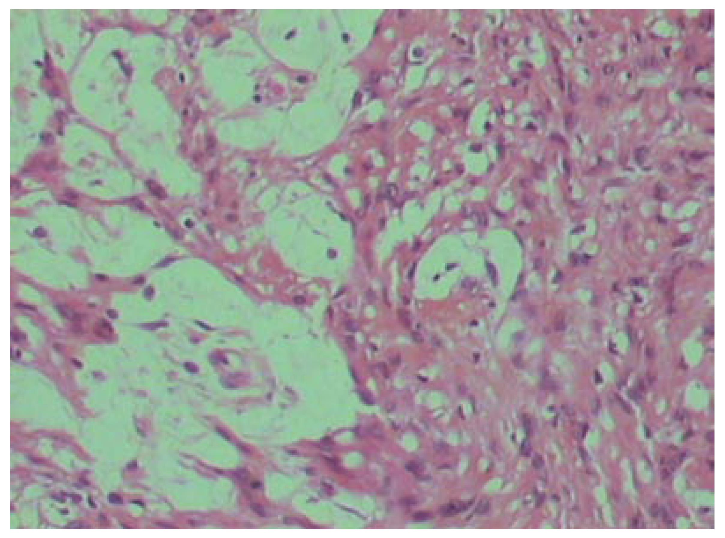
Figure 5 Antoni A area on the right (well-organised spindle cells in a palisade pattern) and Antoni B area (less cellular, loose pleomorphic cells) on the left (HE, × 200).
- Citation: Fass G, Hossey D, Nyst M, Smets D, Saligheh EN, Duttmann R, Claes K, da Costa PM. Benign retroperitoneal schwannoma presenting as colitis: A case report. World J Gastroenterol 2007; 13(41): 5521-5524
- URL: https://www.wjgnet.com/1007-9327/full/v13/i41/5521.htm
- DOI: https://dx.doi.org/10.3748/wjg.v13.i41.5521


