Copyright
©2007 Baishideng Publishing Group Inc.
World J Gastroenterol. Oct 28, 2007; 13(40): 5391-5393
Published online Oct 28, 2007. doi: 10.3748/wjg.v13.i40.5391
Published online Oct 28, 2007. doi: 10.3748/wjg.v13.i40.5391
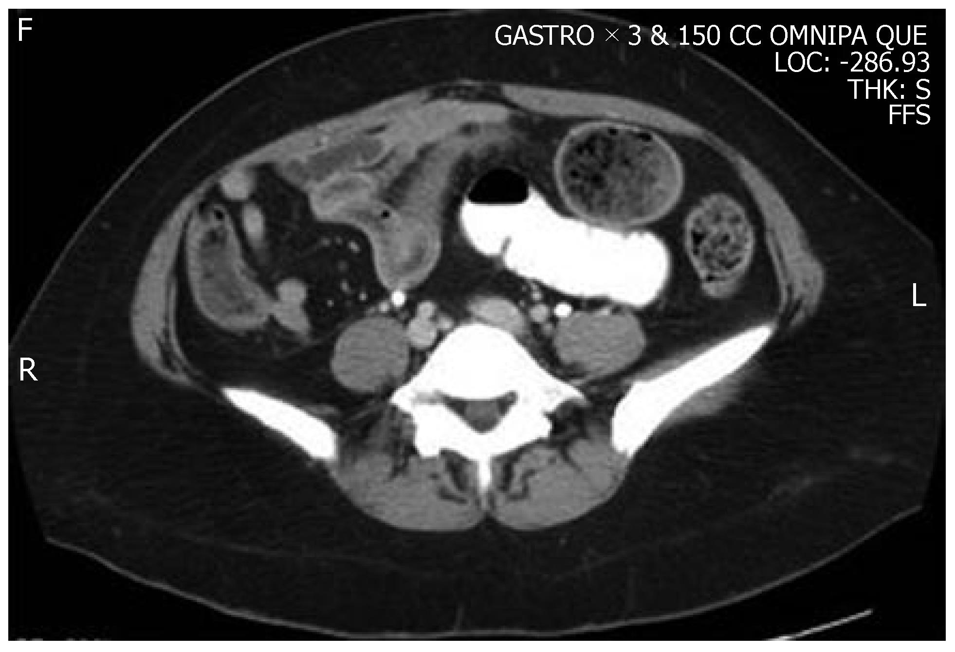
Figure 1 CT scan of the abdomen.
A CT scan of the abdomen revealed an SBO with dilation of small-intestinal loops, and a transition point in the mid-jejunum. A distended small was observed anteriorly of the air fluid level, and distal to this, a dilated loop and cross section was filled with feces, the so-called small bowel feces sign, which is indicative of SBO. Distal to the obstruction, there was a clear transition point in the mid-jejunum.
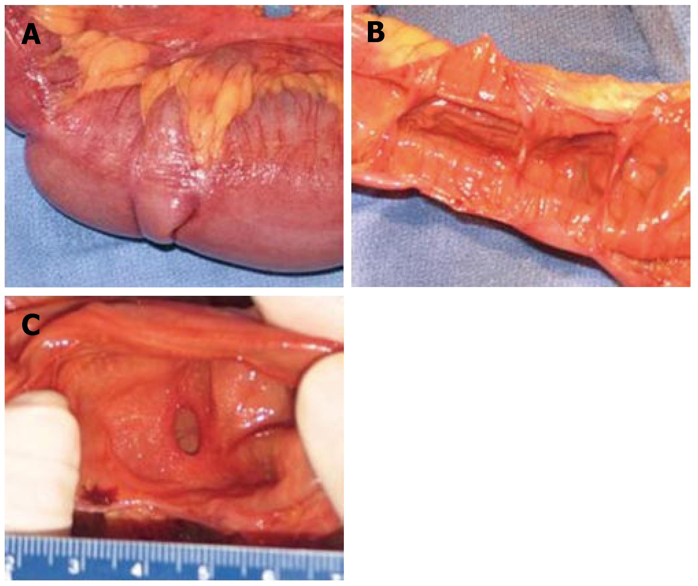
Figure 2 Gross specimens.
Seventy-six centimeter segment of small intestine. On gross examination, fat surrounded the serosal surface of the intestine at irregular intervals, at the mesenteric border (A). Associated with these deposits of fat were 14 areas of mucosal stricture. In the normal intestine, the luminal diameter ranged from 4.7 to 9.5 cm. In the areas of luminal constriction, the diameter was 0.7 to 1.0 cm (B, C).
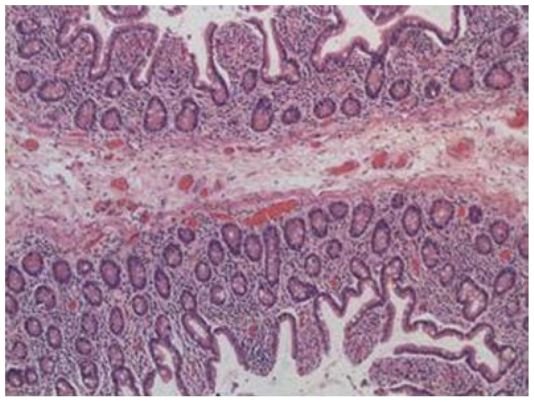
Figure 3 Microscopic image.
On microscopic examination, the intestinal webs contained mucosa and submucosa. Muscularis propria was not present within the webs. There was no evidence of inflammatory bowel disease, malignancy, or pathologic inflammation of the bowel or mesentery.
- Citation: Van Buren II G, Teichgraeber DC, Ghorbani RP, Souchon EA. Sequential stenotic strictures of the small bowel leading to obstruction. World J Gastroenterol 2007; 13(40): 5391-5393
- URL: https://www.wjgnet.com/1007-9327/full/v13/i40/5391.htm
- DOI: https://dx.doi.org/10.3748/wjg.v13.i40.5391


