Copyright
©2007 Baishideng Publishing Group Co.
World J Gastroenterol. Oct 14, 2007; 13(38): 5158-5163
Published online Oct 14, 2007. doi: 10.3748/wjg.v13.i38.5158
Published online Oct 14, 2007. doi: 10.3748/wjg.v13.i38.5158
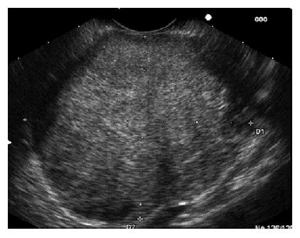
Figure 1 Endoscopic ultrasound image (EUS) showing a mass in body and tail of the pancreas.
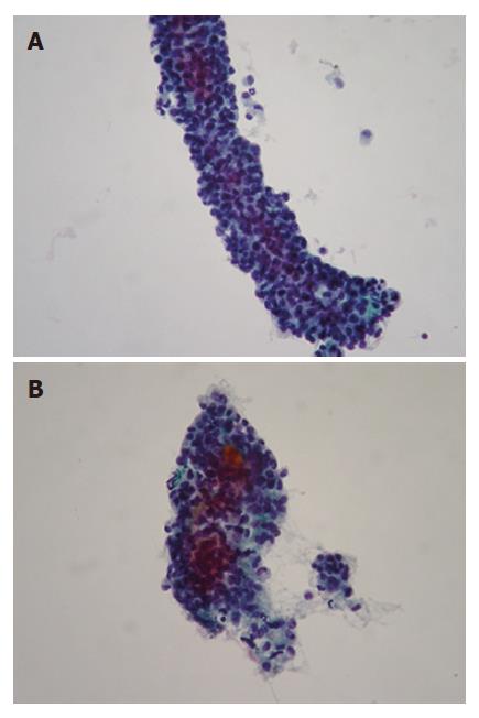
Figure 2 Papillary arrangement composed of delicate fibrovascular core with attached monotonous cuboidal neoplastic cells (A) and adenoid structures composed of cuboidal neoplastic cells (B) (PAP, x 400).
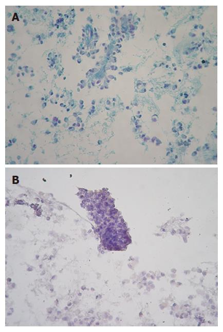
Figure 3 Staining with periodic acid Schiff (PAS) (A) and Alcian-blue (AB) (B) (× 400).
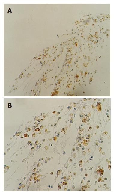
Figure 4 Immunostain for vimentin (A) and CA 19.
9 (B) (x 400).
- Citation: Salla C, Chatzipantelis P, Konstantinou P, Karoumpalis I, Pantazopoulou A, Dappola V. Endoscopic ultrasound-guided fine-needle aspiration cytology diagnosis of solid pseudopapillary tumor of the pancreas: A case report and literature review. World J Gastroenterol 2007; 13(38): 5158-5163
- URL: https://www.wjgnet.com/1007-9327/full/v13/i38/5158.htm
- DOI: https://dx.doi.org/10.3748/wjg.v13.i38.5158


