©2007 Baishideng Publishing Group Co.
World J Gastroenterol. Sep 7, 2007; 13(33): 4437-4444
Published online Sep 7, 2007. doi: 10.3748/wjg.v13.i33.4437
Published online Sep 7, 2007. doi: 10.3748/wjg.v13.i33.4437
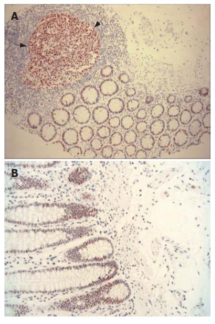
Figure 1 MLH1 and MSH2 expression.
A: Nuclear MLH1 expression detected in germinal centre of lymphoid follicule (dark arrows) and in epithelia of normal colonic mucosa (× 100); B: Crypt epithelia showing normal positive nuclear staining with MSH2 (× 200).
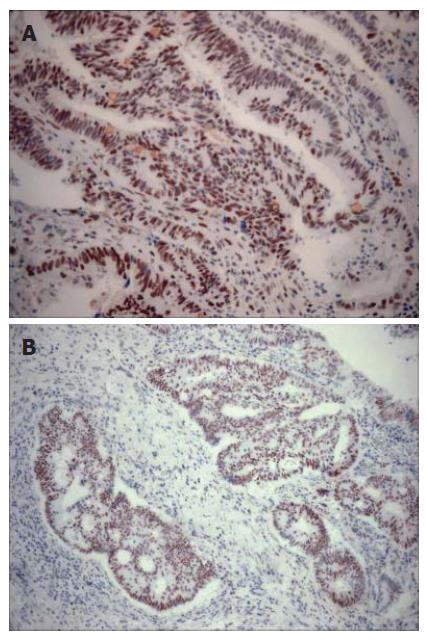
Figure 2 MLH1 and MSH2 expression.
A: Extensive nuclear staining with MLH1 in adenocarcinoma of colon (× 200); B: Tumor cells showing strong positive nuclear staining with MSH2 (× 100).
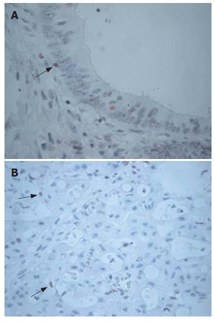
Figure 3 Loss of MLH1 and MSH2 expression in colorectal cancer.
A: Loss of staining with MLH1 in cancer cells, although lymphocytes (arrow) show positive staining (× 400); B: Adenocarcinoma with complete loss of MSH2 expression. Nuclear staining of lymphocytes (arrows) in the stroma served as internal positive control (× 200).
- Citation: Erdamar S, Ucaryilmaz E, Demir G, Karahasanoglu T, Dogusoy G, Dirican A, Goksel S. Importance of MutL homologue MLH1 and MutS homologue MSH2 expression in Turkish patients with sporadic colorectal cancer. World J Gastroenterol 2007; 13(33): 4437-4444
- URL: https://www.wjgnet.com/1007-9327/full/v13/i33/4437.htm
- DOI: https://dx.doi.org/10.3748/wjg.v13.i33.4437


