Copyright
©2007 Baishideng Publishing Group Co.
World J Gastroenterol. Aug 21, 2007; 13(31): 4177-4184
Published online Aug 21, 2007. doi: 10.3748/wjg.v13.i31.4177
Published online Aug 21, 2007. doi: 10.3748/wjg.v13.i31.4177
Figure 1 Examples of imaging techniques.
A: The MPR (coronal oblique) image shows the extrahepatic bile duct as a hypoattenuated structure (arrows); B: The curved MPR image shows the dilated and tortuous pancreatic duct with a stricture at the distal end (long arrow) in one plane; C: On the MinIP (coronal oblique 6 mm thickness slab) image, the extrahepatic bile duct (arrows) and distal pancreatic duct (arrowheads) are well delineated as hypoattenuated tubular structures.
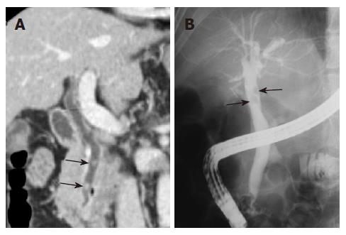
Figure 2 Common bile duct stones in a 65-year-old man with right upper quadrant pain.
A: The MPR (coronal oblique) image demonstrates two stones (arrows) in the common bile duct. B: ERCP shows two round filling defects (arrows) in the extrahepatic bile duct, suggesting stones.
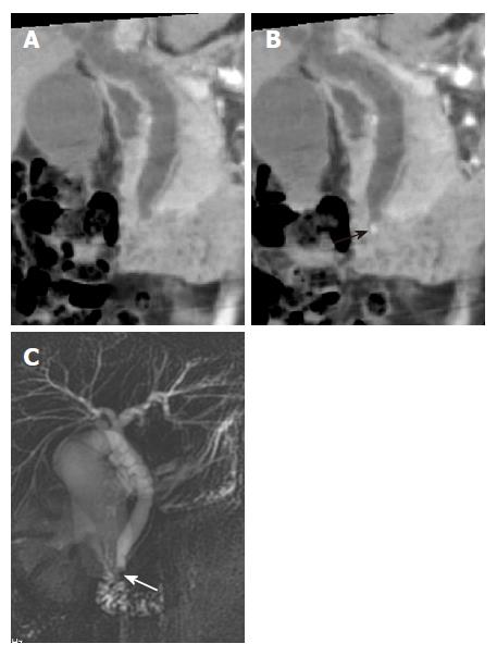
Figure 3 A small stone in the common bile duct of a 34-year-old woman with epigastric pain.
A: MinIP (coronal oblique 6.2 mm thickness slab) image shows mild dilatation of the extrahepatic bile duct without delineation of the stone; B: MinIP (coronal oblique 3.8 mm thickness slab) image with a slab thickness that is thinner than the size of the stone shows the stone (black arrow) in the distal common bile duct; C: MRCP demonstrates the low signal intensity of the stone (white arrow) in the distal common bile duct.
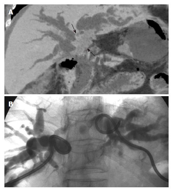
Figure 4 Klatskin tumor in a 68 years old woman with jaundice and fever.
A: MinIP (coronal oblique 14.7 mm thickness slab) image shows an irregular mass (arrows) in the hepatic hilum separating the right and left intrahepatic bile ducts; B: PTC performed by injecting through catheters placed separately shows separation of the right and left intrahepatic bile ducts.
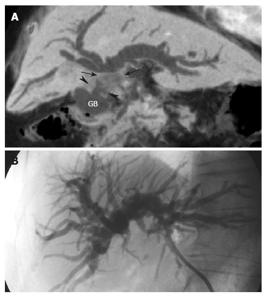
Figure 5 Extrinsic invasion of the common hepatic duct by gallbladder carcinoma in an 80 years old woman with jaundice.
A: MinIP (coronal oblique 14.3 mm thickness slab) image shows dilatation of the intrahepatic bile duct caused by an irregular mass (arrows) in the common hepatic duct, which is the result of direct invasion from the irregular wall thickening (arrowheads) of the gallbladder (GB). This image depicts well the relationship between gallbladder carcinoma and the obstructed biliary duct; B: PTC demonstrates only the biliary obstruction without any suggestion of the cause.
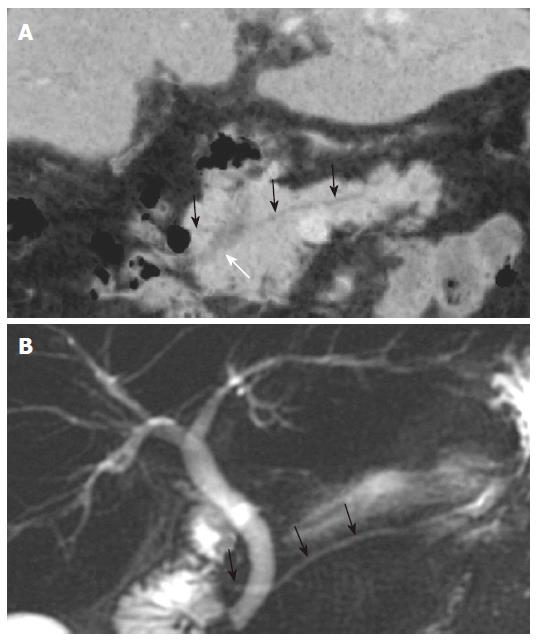
Figure 6 Chronic pancreatitis in a 68 years old woman with epigastric pain.
A: MPR (axial blique) images show an intraductal calculus and stricture (white arrow) with upstream pancreatic duct dilatation (black arrows); B: MRCP demonstrates findings similar to those seen in figure 6A. This diagnosis of chronic pancreatitis was confirmed at surgery.
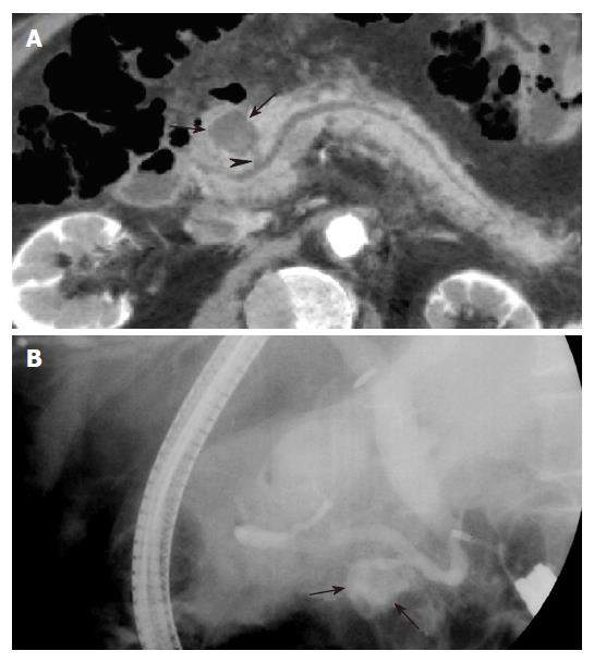
Figure 7 Pancreatic pseudocyst in a 64 years old man with epigastric pain.
A: MinIP (axial oblique 13.8 mm thickness slab) image shows a round cystic lesion (arrows) in the pancreatic head, communicating (arrowhead) with the pancreatic duct. This patient was diagnosed with pancreatitis on clinical and radiological findings (not shown); B: ERCP image obtained with the patient on the right lateral decubitus position shows the cystic lesion (arrows) filled with contrast material, which represents communication between the cyst and the pancreatic duct.
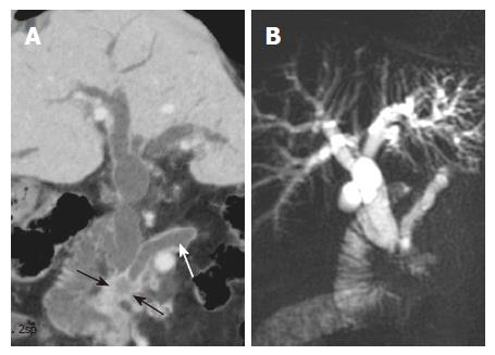
Figure 8 Pancreatic head cancer in a 77 years old woman with jaundice.
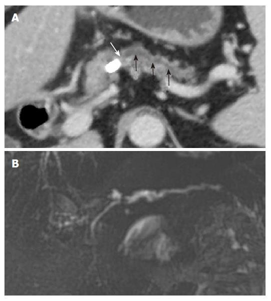
A: MinIP (coronal oblique 6.1 mm thickness slab) image demonstrates dilatation of both the bile duct and the pancreatic duct (white arrow), caused by cancer (black arrows) of the pancreatic head; B: MRCP shows dilatation of the bile duct and the pancreatic duct (double duct sign), which is a typical finding in pancreatic head cancer.
Figure 9 Branch duct type of intraductal papillary mucinous neoplasm of the pancreas in a 69 years old woman with epigastric discomfort.
A: MinIP (axial oblique 6.2 mm thickness slab) image shows a lobulated cystic lesion (arrows) that is contiguous with the mildly prominent main pancreatic duct (black arrowheads); B: The curved MPR image shows a communication (white arrow) between the cystic mass and the pancreatic duct; C: ERCP shows a cystic branch duct (black arrows) with an intraluminal filling defect (white arrowhead) that represents mucus. Mucus was seen protruding from a patulous duodenal papilla (not shown).
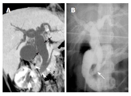
Figure 10 Choledochal cyst in a 7 years old girl with jaundice.
A: MinIP (coronal oblique 14.1 mm thickness slab) image shows dilatation of the entire extrahepatic bile duct (black arrows) and a long common channel (white arrow). Note the subtle increased attenuation (arrowheads) within the distal common bile duct, which represents stones; B: Operative cholangiography shows dilatation of the extrahepatic bile duct with a long common channel (white arrow), which is identical to the findings obtained by MinIP (A).
Figure 11 Pancreatic divisum in a 68-year-old man with epigastric discomfort.
A: MinIP (coronal oblique 6.2 mm thickness slab) images show the dorsal pancreatic duct (black arrows) crossing the distal common bile duct (white arrow) superiorly to enter the minor papilla; B: MRCP shows the prominent dorsal pancreatic duct (black arrows) crossing the distal common bile duct. The ventral duct is not visualized.
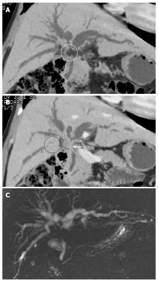
Figure 12 Klatskin’s tumor in a 65 years old man with jaundice.
A: MinIP (coronal oblique 18.8 mm thickness slab) image shows focal disruption (white circle) of the common hepatic duct due to the adjacent perihepatic fat; B: MinIP (coronal oblique 6.2 mm thickness slab) image shows focal loss (black circle) of the right posterior inferior segmental duct due to exclusion by out of the slab range. However, severe narrowing of the common hepatic duct (white arrow) is better visualized than that seen on (A); C: MRCP shows a typical Klatskin tumor with narrowing of the confluent portion of both intrahepatic bile ducts.
- Citation: Kim HC, Yang DM, Jin W, Ryu CW, Ryu JK, Park SI, Park SJ, Shin HC, Kim IY. Multiplanar reformations and minimum intensity projections using multi-detector row CT for assessing anomalies and disorders of the pancreaticobiliary tree. World J Gastroenterol 2007; 13(31): 4177-4184
- URL: https://www.wjgnet.com/1007-9327/full/v13/i31/4177.htm
- DOI: https://dx.doi.org/10.3748/wjg.v13.i31.4177


