Copyright
©2007 Baishideng Publishing Group Co.
World J Gastroenterol. Jan 21, 2007; 13(3): 470-473
Published online Jan 21, 2007. doi: 10.3748/wjg.v13.i3.470
Published online Jan 21, 2007. doi: 10.3748/wjg.v13.i3.470
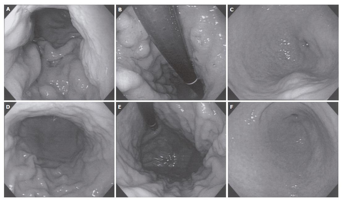
Figure 1 Gastroscopic findings at diagnosis (upper lane, A-C) and after 4 courses of chemotherapy (lower lane, D-F).
A: Lower body: Wall thickening and erosions can be observed; B: Mid body: Wall expandability is extremely poor in this part; C: Antrum: An SMT like lesion at the greater curvature suggests submucosal invasion of the cancer; D: Lower body: Wall thickening is less prominent; E: Mid body: Wall expandability has drastically improved; F: Antrum: the SMT like lesion has disappeared.
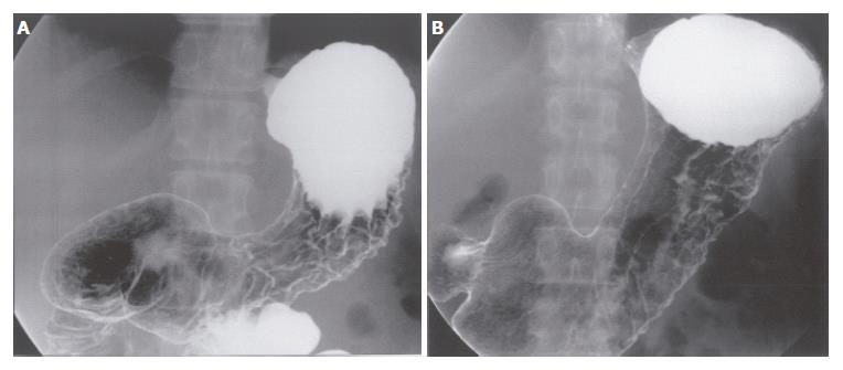
Figure 2 Upper gastrointestinal series before (A) and after treatment (B).
A: At diagnosis, wall thickening and loss of expandability is prominent at the body, while normal mucosa is relatively spared at the antrum; B: After 6 courses of chemotherapy, the expandability at the body has restored.
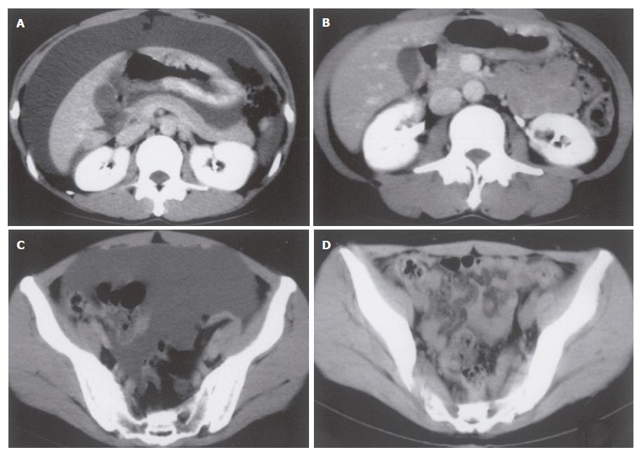
Figure 3 CT scan findings before (Left, A and C) and after 6 courses of chemotherapy (Right, B and D).
A: Antral level: Wall thickening at the body and massive ascites are detected; B: Antral level: Wall thickening at the body is less prominent and no ascites can be observed; C: Pelvic level: Massive ascites extend to pelvic level; D: Pelvic level: Ascites have completely vanished.
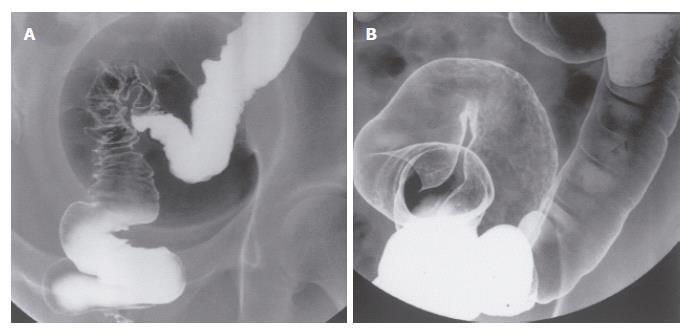
Figure 4 Barium enema before (A) and after treatment (B).
A: At diagnosis, poorly expandable segments within the rectosigmoid suggest severe malignant ascites; B: After 6 courses of chemotherapy, the rectosigmoidal colon can fully expand with air enema.
- Citation: Koizumi Y, Obata H, Hara A, Nishimura T, Sakamoto K, Fujiyama Y. A case of scirrhous gastric cancer with peritonitis carcinomatosa controlled by TS-1® + paclitaxel for 36 mo after diagnosis. World J Gastroenterol 2007; 13(3): 470-473
- URL: https://www.wjgnet.com/1007-9327/full/v13/i3/470.htm
- DOI: https://dx.doi.org/10.3748/wjg.v13.i3.470


