Copyright
©2007 Baishideng Publishing Group Co.
World J Gastroenterol. Jan 21, 2007; 13(3): 341-348
Published online Jan 21, 2007. doi: 10.3748/wjg.v13.i3.341
Published online Jan 21, 2007. doi: 10.3748/wjg.v13.i3.341
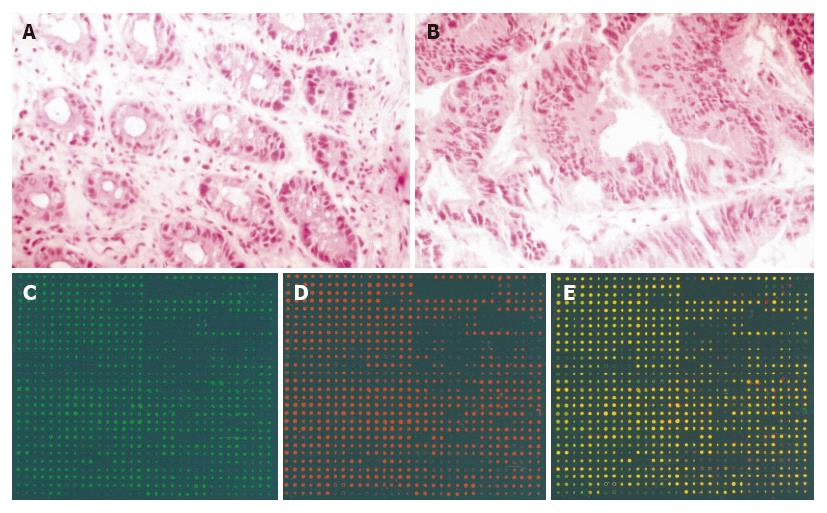
Figure 1 Histopathological examination of the tumor tissues of rectal cancer.
A: Normal rectal mucosa tissue; B: Adenocarcinoma of rectal tissues; C, D and E: images of normal tissue, tumor tissues and overlay image of the tumor to normal of patient No.19 respectively. The density of the spot reflects the fluorescent intensity of the genes. In the overlay images, the yellow spots represent the expression of the genes in tumor tissues and normal tissues were not significant. The red spots represent the up-regulated genes in rectal cancer tissues. The green spots represent the down-regulated genes in rectal cancer tissues.
Figure 2 Semi-quantitative RT-PCR of MTA1 mRNA.
A: Electrophoresis image of MTA1 expression, lanes 1-8 represent the mRNA expression of MTA1 in 4 pairs of rectal cancer patients, respectively. Lanes 1, 3, 5, 7 were normal tissues and 2, 3, 6, 8 were tumor tissues. B: The semi-quantitative analysis results with spot intensity software. The ratios of MTA1 in tumor tissues were lower than that of paired normal tissues.
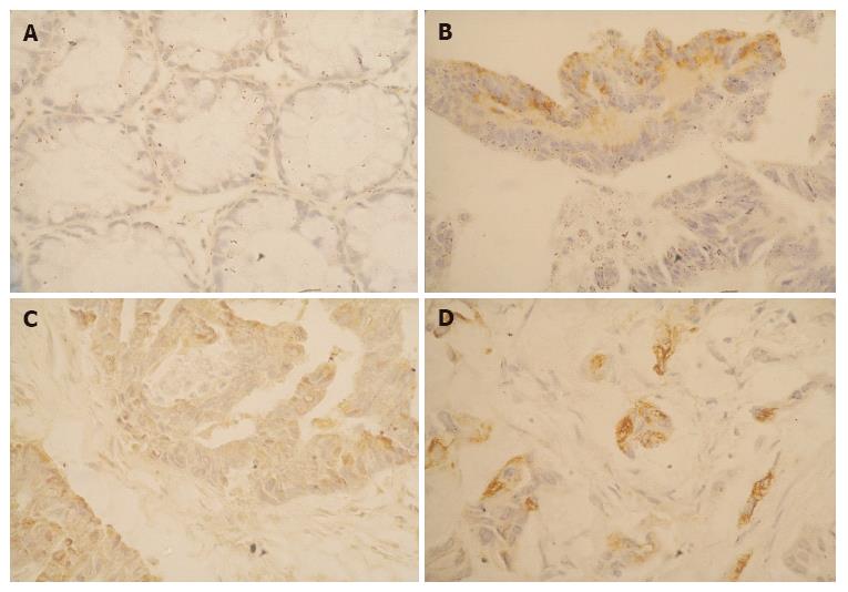
Figure 3 Validation of the expression of BCL-2 with immunohistochemistry method.
A: The normal rectal mucosa. There was no expression of BCL-2 in all the cells; B: The expression of BCL-2 in well differentiated rectal cancer. The tumor cells were stained yellow-brown by DAB; C: The expression of BCL-2 in another intermediately differentiated rectal cancer. The expression of BCL-2 was also significant in tumor cells; D: The expression of BCL-2 in poorly differentiated rectal tissues.
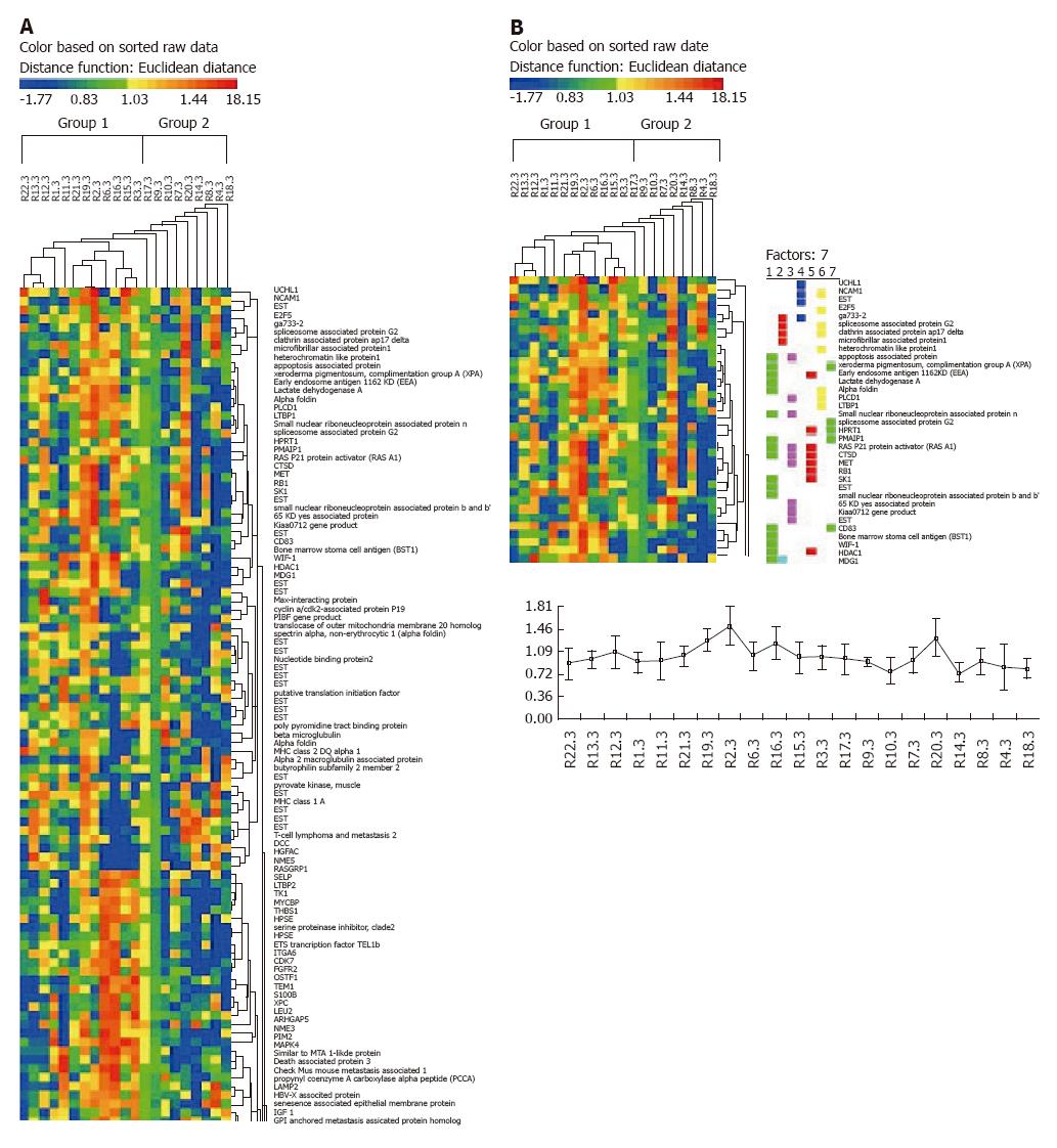
Figure 4 Hierarchial cluster analysis and principal component analysis results.
A: Hierachial cluster analysis of patients and genes. The patients could be classified into two groups: one group of clinicopathological stage above grade II and one group below grade II. B: Principal component analysis of the genes related to rectal cancer.
- Citation: Gao XQ, Han JX, Xu ZF, Zhang WD, Zhang HN, Huang HY. Identification of the differential expressive tumor associated genes in rectal cancers by cDNA microarray. World J Gastroenterol 2007; 13(3): 341-348
- URL: https://www.wjgnet.com/1007-9327/full/v13/i3/341.htm
- DOI: https://dx.doi.org/10.3748/wjg.v13.i3.341


