Copyright
©2007 Baishideng Publishing Group Co.
World J Gastroenterol. Jul 28, 2007; 13(28): 3792-3798
Published online Jul 28, 2007. doi: 10.3748/wjg.v13.i28.3792
Published online Jul 28, 2007. doi: 10.3748/wjg.v13.i28.3792
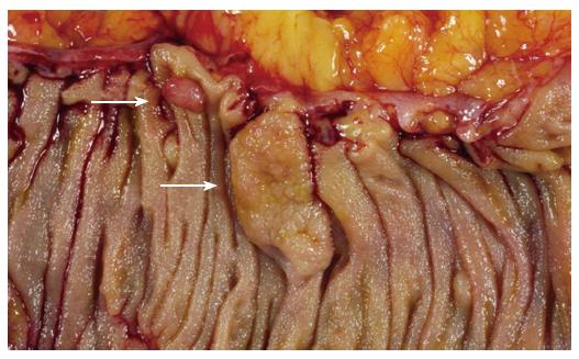
Figure 1 Sessile serrated adenoma (lower arrow).
These polyps tend to be large, sessile and situated on the crest of the mucosal folds. This appearance contrasts with that of a typical adenomatous polyp (upper arrow).
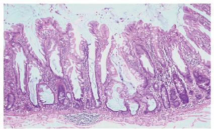
Figure 2 Low power appearance of a sessile serrated adenoma.
Serration is seen to the base of the crypts, which are dilated and may show growth parallel to the muscularis mucosae (HE, x 20).
Figure 3 The crypts of a sessile serrated adenoma may herniate through the muscularis mucosae, producing an appearance which may be confused with invasive tumour (HE, x 20).
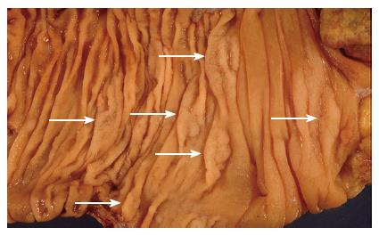
Figure 4 An example of hyperplastic polyposis, demonstrating numerous sessile serrated adenomas (arrows).
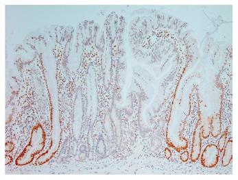
Figure 5 Immunohistochemical staining for the mismatch repair protein MLH1, demonstrating “clonal” loss of expression in a sessile serrated adenoma (x 20).
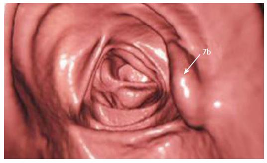
Figure 6 Virtual colonoscopic image demonstrating a sessile serrated adenoma (arrow).
- Citation: Harvey NT, Ruszkiewicz A. Serrated neoplasia of the colorectum. World J Gastroenterol 2007; 13(28): 3792-3798
- URL: https://www.wjgnet.com/1007-9327/full/v13/i28/3792.htm
- DOI: https://dx.doi.org/10.3748/wjg.v13.i28.3792


