©2007 Baishideng Publishing Group Inc.
World J Gastroenterol. Jul 14, 2007; 13(26): 3641-3644
Published online Jul 14, 2007. doi: 10.3748/wjg.v13.i26.3641
Published online Jul 14, 2007. doi: 10.3748/wjg.v13.i26.3641
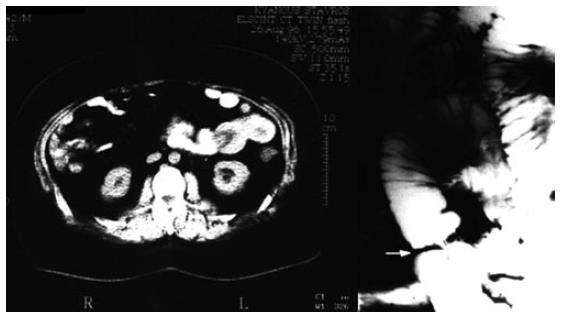
Figure 1 Abdominal computed tomography showed an obstructive lesion of the jejunum with dilatation of the edematous adjacent bowel loops.
Enteroclysis showed a circular obstructive lesion of the jejunum with apple-core appearance. Close observation of the lesion shows the presence of radiation-lucent linear markings, with barium fillings on either side, projecting to the adjacent loop consistent with the walls of the intussusceptum.
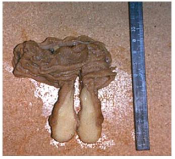
Figure 2 A pedunculated lesion, measuring 4 cm × 4 cm, was the lead point of the intussusception.
The lesion was covered with mucosa and was composed of smooth submucosal fat. The clinical diagnosis of jejunal lipoma was subsequently confirmed at pathologic evaluation.
- Citation: Manouras A, Lagoudianakis EE, Dardamanis D, Tsekouras DK, Markogiannakis H, Genetzakis M, Pararas N, Papadima A, Triantafillou C, Katergiannakis V. Lipoma induced jejunojejunal intussusception. World J Gastroenterol 2007; 13(26): 3641-3644
- URL: https://www.wjgnet.com/1007-9327/full/v13/i26/3641.htm
- DOI: https://dx.doi.org/10.3748/wjg.v13.i26.3641


