©2007 Baishideng Publishing Group Inc.
World J Gastroenterol. Jul 7, 2007; 13(25): 3466-3471
Published online Jul 7, 2007. doi: 10.3748/wjg.v13.i25.3466
Published online Jul 7, 2007. doi: 10.3748/wjg.v13.i25.3466
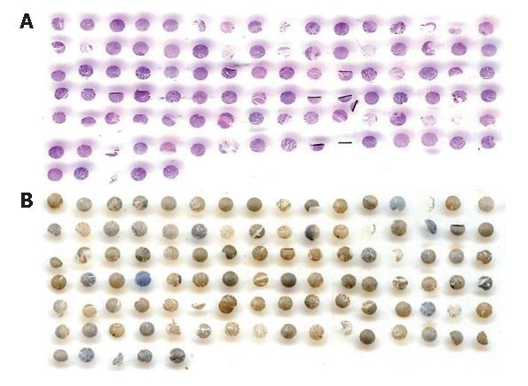
Figure 1 Scanning image of tissue array.
A: HE Staining; B: COX-2 immuno-histochemical staining.
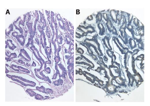
Figure 2 Scanning image of tissue array.
A: HE, × 100; B: COX-2 immuno-histochemical staining, × 100.
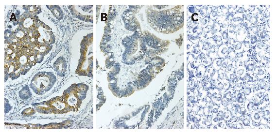
Figure 3 COX-2 expression in gastric cancer.
A: Positive COX-2 expression in the well-differentiated adenocarcinoma (Envision, × 200); B: Positive COX-2 expression in the moderately-differentiated adenocarcinoma (Envision, × 200); C: Negative COX-2 expression in the normal gastric mucosa (Envision, × 200).
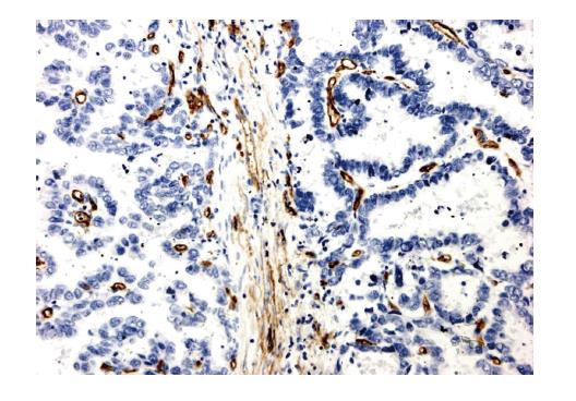
Figure 4 CD34 expression in the gastric tissue.
Immunostaining of CD34 in the cytoplasm and membrane of endothelial cells (Envision, × 200).
- Citation: Mao XY, Wang XG, Lv XJ, Xu L, Han CB. COX-2 expression in gastric cancer and its relationship with angiogenesis using tissue microarray. World J Gastroenterol 2007; 13(25): 3466-3471
- URL: https://www.wjgnet.com/1007-9327/full/v13/i25/3466.htm
- DOI: https://dx.doi.org/10.3748/wjg.v13.i25.3466


