©2007 Baishideng Publishing Group Co.
World J Gastroenterol. Jun 21, 2007; 13(23): 3265-3267
Published online Jun 21, 2007. doi: 10.3748/wjg.v13.i23.3265
Published online Jun 21, 2007. doi: 10.3748/wjg.v13.i23.3265
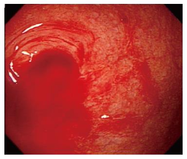
Figure 1 Continuous oozing of fresh blood is seen from the appendiceal orifice.
Figure 2 Small diverticula without evidence of active bleeding are found in the cecum.
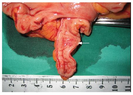
Figure 3 The inside of the appendiceal specimen appears normal, but a small nodule is found in the mid portion (white arrow).
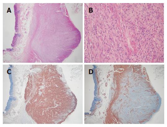
Figure 4 A: Whole layers of appendix show an ill-defined mass involving the proper muscle layer (HE, × 20); B: The mass is composed of spindle cells usually in fascicular pattern.
The neoplastic cells are characterized by spindle nuclei with blunt or tapered end and bland chromatin pattern and by various amounts of amphophilic cytoplasm and ill-defined cellular borders (HE, × 200); C: The neoplastic cells show immunopositive staining for CD34 (Immunohistochemical stain, × 20); D: The neoplastic cells show immunonegative staining for smooth muscle actin, but positive in the remaining proper muscle cells (Immunohistochemical stain, × 20).
- Citation: Kim KJ, Moon W, Park MI, Park SJ, Lee SH, Chun BK. Gastrointestinal stromal tumor of appendix incidentally diagnosed by appendiceal hemorrhage. World J Gastroenterol 2007; 13(23): 3265-3267
- URL: https://www.wjgnet.com/1007-9327/full/v13/i23/3265.htm
- DOI: https://dx.doi.org/10.3748/wjg.v13.i23.3265


