©2007 Baishideng Publishing Group Co.
World J Gastroenterol. Jun 14, 2007; 13(22): 3095-3100
Published online Jun 14, 2007. doi: 10.3748/wjg.v13.i22.3095
Published online Jun 14, 2007. doi: 10.3748/wjg.v13.i22.3095
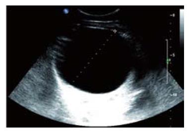
Figure 1 Abdominal ultrasound of a solitary simple liver cyst showing a well-defined anechoic unilocular fluid-filled lesion with posterior acoustic enhancement.
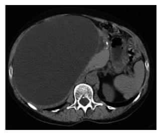
Figure 2 Abdominal com-puted tomography of a large solitary simple liver cyst showing a well-demarcated lesion with fluid density and without enhancement after contrast administration.
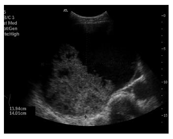
Figure 3 Abdominal ultrasound of a complicated liver cyst showing a well defined hypoechogenic lesion with solid appearing blood clot contents.
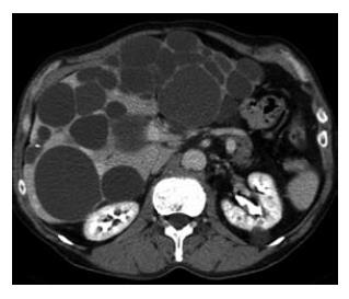
Figure 4 Abdominal computed tomography of a patient with PLD showing multiple cysts throughout the liver.
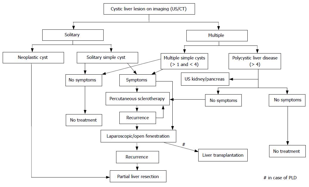
Figure 5 Proposed algorythm in the management of patients with cystic liver lesions.
US indicates ultrasound; CT: computed tomography; PLD: polycystic liver disease.
- Citation: Erdogan D, van Delden OM, Rauws EA, Busch OR, Lameris JS, Gouma DJ, van Gulik TM. Results of percutaneous sclerotherapy and surgical treatment in patients with symptomatic simple liver cysts and polycystic liver disease. World J Gastroenterol 2007; 13(22): 3095-3100
- URL: https://www.wjgnet.com/1007-9327/full/v13/i22/3095.htm
- DOI: https://dx.doi.org/10.3748/wjg.v13.i22.3095


