©2007 Baishideng Publishing Group Co.
World J Gastroenterol. May 28, 2007; 13(20): 2892-2894
Published online May 28, 2007. doi: 10.3748/wjg.v13.i20.2892
Published online May 28, 2007. doi: 10.3748/wjg.v13.i20.2892
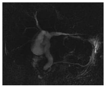
Figure 1 MRCP showing a diffuse dilated common bile duct without signal void and the remarkable pancreatic duct.
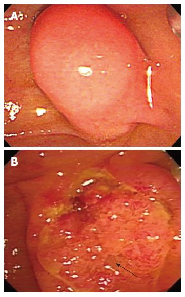
Figure 2 Duodeno-scopy showing a bulged major papilla with overlying intact mucosa (A) and an even and firm nodular mass with mucosal and villous granularities origionating from the peripancreatic orifice after endoscopic biliary sphinctertomy (B) (the arrow indicates the main pancreatic duct orifice).
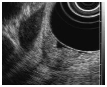
Figure 3 EUS showing slightly elevated echo-genic lesions on major papilla.
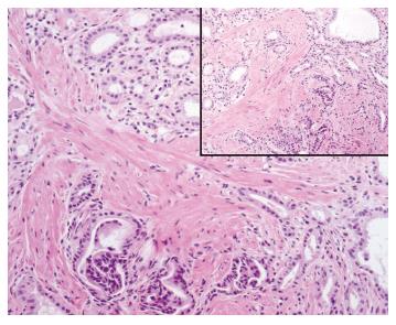
Figure 4 Histology of the resected spe-cimen showing nu-merous ductules in association with promi-nent proliferation of smooth muscle in the submucosa of the duodenum (insert, HE, × 2).
The ductules were lined by columnar epithelial cells (HE, × 200).
- Citation: Kwon TH, Park DH, Shim KY, Cho HD, Park JH, Lee SH, Chung IK, Kim HS, Park SH, Kim SJ. Ampullary adenomyoma presenting as acute recurrent pancreatitis. World J Gastroenterol 2007; 13(20): 2892-2894
- URL: https://www.wjgnet.com/1007-9327/full/v13/i20/2892.htm
- DOI: https://dx.doi.org/10.3748/wjg.v13.i20.2892


