©2007 Baishideng Publishing Group Co.
World J Gastroenterol. May 21, 2007; 13(19): 2707-2716
Published online May 21, 2007. doi: 10.3748/wjg.v13.i19.2707
Published online May 21, 2007. doi: 10.3748/wjg.v13.i19.2707
Figure 1 A representative two-color flow cytometric analysis for human cell engraftment in PB of human-rat chimeras.
After birth, at the indicated time points, peripheral blood (PB) was collected, and subsequently the collected blood cells were stained with both anti-human CD45 and anti-human CD20 antibodies and analyzed with flow cytometry. Human PB from healthy volunteers was employed as positive control (PC). Negative control (NC): normal rat PB.
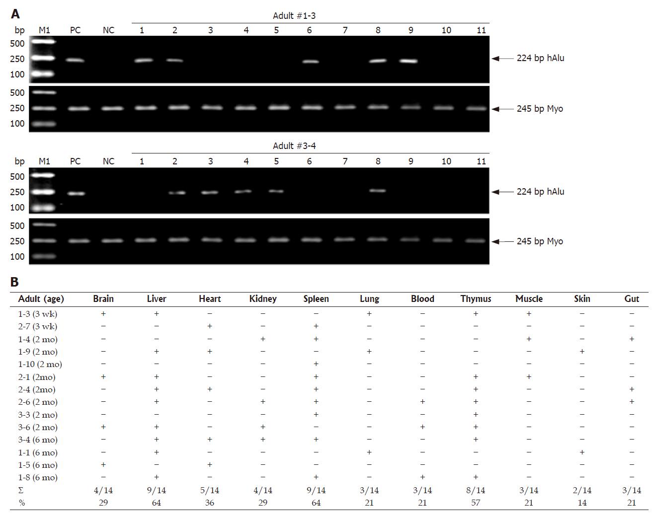
Figure 2 Human donor contributions in different adult tissues analyzed by human gene-specific PCR.
A: Identification of human-specific gene in different adult tissues of MNC-transplanted rats. To determine human donor contribution in MNC-transplanted rats, PCR was carried out on genomic DNA with primers specific for hAlu gene (Table 1). Two typical analyses of PCR products by agarose gel electrophoresis from positive-control human blood genomic DNA (lane PC), normal/non-transplanted rat genomic DNA (lane NC) and genomic DNA prepared from different adult tissues, i.e. brain (lane 1), liver (lane 2), heart (lane 3), kidney (lane 4), spleen (lane 5), lung (lane 6), PB (lane 7), thymus (lane 8), muscle (lane 9), skin (lane 10) and gut (lane 11), of two MNC-transplanted rats are shown. Myogenin (Myo)-specific PCRs were carried out as for quality and quantity controls of genomic DNA. Data are representative of three independent PCRs that yielded similar results. The arrow indicates the position of PCR products amplified by the primers shown in Table 1. M1: Molecular weight DNA marker (DL2000, TaKaRa). Other details are as in Figure 1 and Table 2; B: Summary of tissue distribution of human cells in different tissues of MNC-transplanted adult rats. The total fraction (Σ) of all adults with positive signals in a specific tissue is summarized below the respective column and also shown as a percentage (%) to enable comparisons. –: No donor signal; +: Positive donor signal.
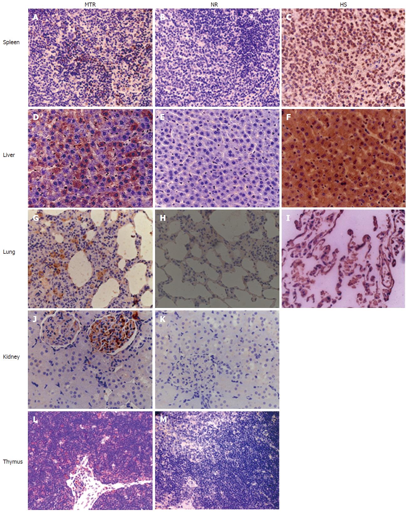
Figure 3 Immunohistochemical analysis for human β2-microglobulin antigen in various tissues of MNC-transplanted rats.
Human β2-microglobulin-positive cells were identified in multiple tissues of MNC-transplanted rats by immunohistochemistry (IHC). Brown staining shows positive human cells in various tissues of MNC-transplanted rats (MTR) and human samples (HS), whereas no positive human cells were found in normal control rats (NR).
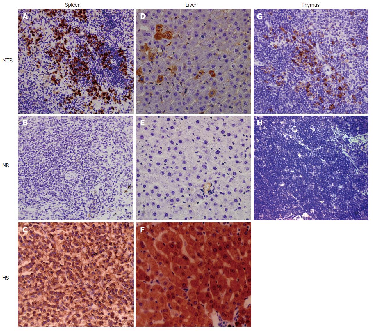
Figure 4 Evidences for site-specific human cell differentiation in the xenogeneic system.
Transplanted UCB-derived human cells underwent site-specific differentiation in various tissues [i.e. liver, spleen and thymus] of MNC-transplanted rats, as confirmed by using human-specific differentiation markers (such as CK18 and CD45). Brown staining shows CD45-positive human cells in the spleen (A and C) and thymus (G) of MNC-transplanted rats (MTR) and human samples (HS), whereas no positive human cells are found in the spleen (B) and thymus (H) of normal control rats (NR). Livers (D and F) from MTR and HS are uniformly positive for CK18, while liver (E) from NR is negative for CK18.
-
Citation: Sun Y, Xiao D, Pan XH, Zhang RS, Cui GH, Chen XG. Generation of human/rat xenograft animal model for the study of human donor stem cell behaviors
in vivo . World J Gastroenterol 2007; 13(19): 2707-2716 - URL: https://www.wjgnet.com/1007-9327/full/v13/i19/2707.htm
- DOI: https://dx.doi.org/10.3748/wjg.v13.i19.2707


