©2007 Baishideng Publishing Group Co.
World J Gastroenterol. May 21, 2007; 13(19): 2684-2692
Published online May 21, 2007. doi: 10.3748/wjg.v13.i19.2684
Published online May 21, 2007. doi: 10.3748/wjg.v13.i19.2684
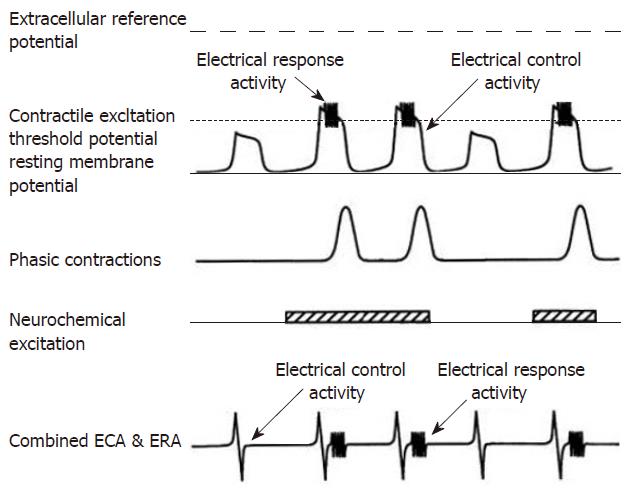
Figure 1 The top electrical tracing illustrates the relationship of intracellularly recorded myoelectric activity and neurochemical excitation with contractions.
In the absence of neurochemical excitation, the ECA depolarizations do not exceed the contractile excitation threshold potential. Consequently, no ERA burst is recorded and no contraction occurs during the first and fourth depolarization. When neurochemical stimulation occurs, ECA depolarization exceeds the excitation threshold, an ERA burst occurs and the smooth muscle contracts (cycle 2, 3 and 5). The extracellular electrode recordings are shown in the last tracing and record the follow of currents from a large numbers of smooth muscle cells. The relationship between ECA, neurochemical excitation ERA and contractions is the same as with intracellular recordings. (From Sarna, SK: In vivo myoelectric activity: Methods, analysis and interpretation. In Wood JD (ed): Handbook of Physiology: A Critical, Comprehensive Presentation of Physiological Knowledge and Concepts. Bethesda, MD, American Physiological Society, 1989: 817-863).
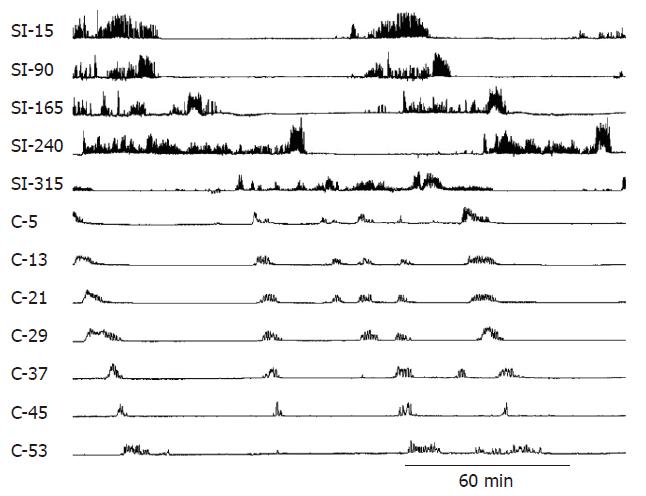
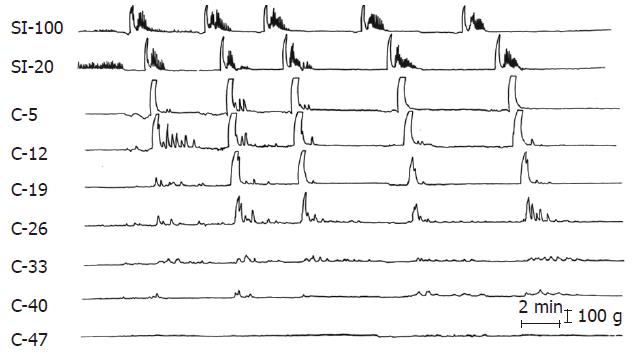
Figure 2 Normal small intestinal and colonic MMCs.
Note how the phasic contractions of the small intestine have a shorter duration and different character from the contractions of the colon. The numbers on the left-side indicate the distance in centimeters of the small intestine (SI) from the pylorus. The letter C refers to the colon and the numbers are the distance in cm from the ileocolonic junction.
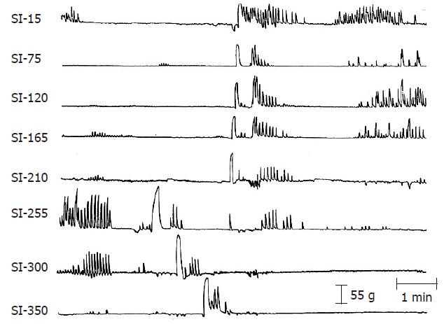
Figure 3 A RGC and a GMC following radiation.
The two giant contractions originated about 255 cm from the pylorus and migrated in opposite directions. The duration of the GMC is longer than that of the RGC but the RGC migrates more rapidly. The numbers on the left-side indicate the distance in centimeters of the small intestine (SI) from the pylorus. (From Otterson MF, Sarna SK, Moulder JE. Effects of fractionated doses of ionizing radiation on small intestinal motor activity. Gastroenterology 1988; 95: 1249-1257.).
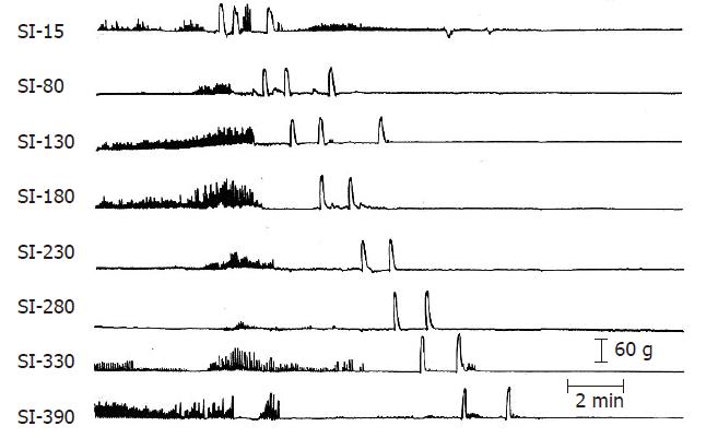
Figure 4 Three GMCs originating in the duodenum following irradiation.
The first two GMCs migrated to the ileocolonic junction but the third stopped at 130 cm from the pylorus. See Figure 3 caption for details. (From Otterson MF, Sarna SK, Moulder JE. Effects of fractionated doses of ionizing radiation on small intestinal motor activity. Gastroenterology 1988; 95: 1249-1257).
Figure 5 Five GMCs originating in the small intestine at greater than 100 cm from the ileocolonic junction are migrating into the proximal colon following radiation.
SI refers to the small intestine; C to colonic strain gauges and the numbers are the distance in cm from the ileocolonic junction. (From Otterson MF et al, Effects of fractionated doses of ionizing radiation on colonic motor activity. Am J Physiol 1992; 263(4 Pt 1): G518-G526.).
- Citation: Otterson MF. Effects of radiation upon gastrointestinal motility. World J Gastroenterol 2007; 13(19): 2684-2692
- URL: https://www.wjgnet.com/1007-9327/full/v13/i19/2684.htm
- DOI: https://dx.doi.org/10.3748/wjg.v13.i19.2684


