Copyright
©2007 Baishideng Publishing Group Co.
World J Gastroenterol. Apr 28, 2007; 13(16): 2319-2323
Published online Apr 28, 2007. doi: 10.3748/wjg.v13.i16.2319
Published online Apr 28, 2007. doi: 10.3748/wjg.v13.i16.2319
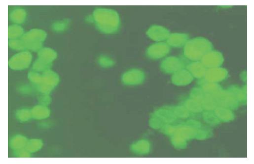
Figure 1 Sorted cells stained with FITC labeled anti human AFP antibody and observed under fluorescent (Zeiss) microscope (× 400).
Cells were observed to be positive for AFP and a thin rim of cytoplasm with a big nucleus.
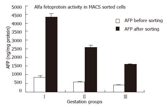
Figure 2 The graph shown is representative of the three independent experiments.
The activity was expressed in μg/mg protein.
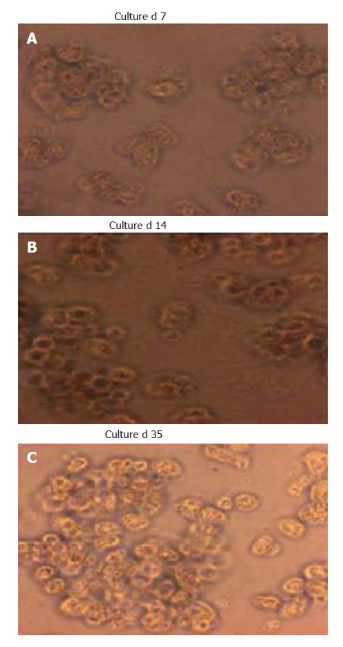
Figure 3 A: The sorted cells on d 0 of culture.
Cells were cultured in the long term. The cells are showing intact morphology (× 400); B: The cultured cells on 14th d. The cells are showing intact morphology (× 400); C: The cultured cells on 35th d. The cells are showing intact morphology (× 400).
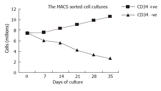
Figure 4 Culture of CD34+ sorted primary cells.
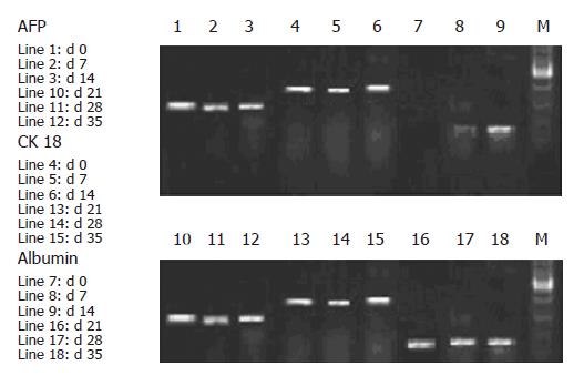
Figure 5 Gene expression analysis in different days of cultures of hepatic progenitors.
- Citation: Nyamath P, Alvi A, Habeeb A, Khosla S, Khan AA, Habibullah C. Characterization of hepatic progenitors from human fetal liver using CD34 as a hepatic progenitor marker. World J Gastroenterol 2007; 13(16): 2319-2323
- URL: https://www.wjgnet.com/1007-9327/full/v13/i16/2319.htm
- DOI: https://dx.doi.org/10.3748/wjg.v13.i16.2319


