Copyright
©2007 Baishideng Publishing Group Co.
World J Gastroenterol. Mar 28, 2007; 13(12): 1877-1878
Published online Mar 28, 2007. doi: 10.3748/wjg.v13.i12.1877
Published online Mar 28, 2007. doi: 10.3748/wjg.v13.i12.1877
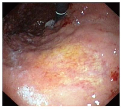
Figure 1 Upper GI endoscopy showing a white-yellowish 3-cm circular area with fine granular appearance at the gastric body (above the angulus).
Figure 2 Photomicrograph of the endoscopic biopsy specimens showing deposition of fibrillar eosinophilic substance (A) (HE, x 10), positive Congo red staining (B) (Congo red, x 10), and high power image showing infiltration of the lamina propria with lymphocytes and polyclonal plasma cells (C) (HE, x 25).
- Citation: Rotondano G, Salerno R, Cipolletta F, Bianco MA, De Gregorio A, Miele R, Prisco A, Garofano ML, Cipolletta L. Localized amyloidosis of the stomach: A case report. World J Gastroenterol 2007; 13(12): 1877-1878
- URL: https://www.wjgnet.com/1007-9327/full/v13/i12/1877.htm
- DOI: https://dx.doi.org/10.3748/wjg.v13.i12.1877


