Copyright
©2006 Baishideng Publishing Group Co.
World J Gastroenterol. Feb 21, 2006; 12(7): 1157-1159
Published online Feb 21, 2006. doi: 10.3748/wjg.v12.i7.1157
Published online Feb 21, 2006. doi: 10.3748/wjg.v12.i7.1157
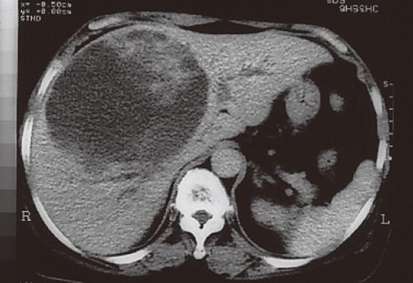
Figure 1 A low-attenuation cystic mass located in segments 4, 5, and 8 with bilateral biliary tract compression in a non-cirrhotic liver by CT.
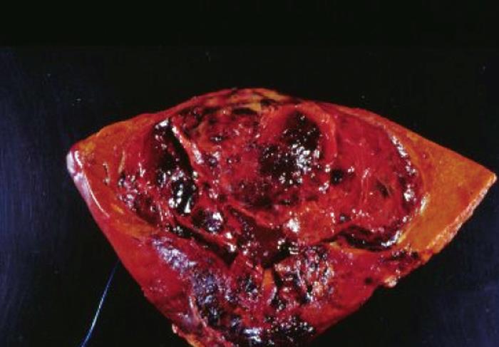
Figure 2 A lobulated liver liposarcoma with intra-tumor hemorrhage.
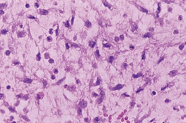
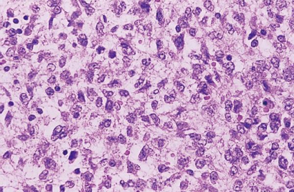
Figure 3 Numerous lipoblasts and capillaries in the myxoid liposarcoma (hematoxylin-eosin staining, 400 ×).
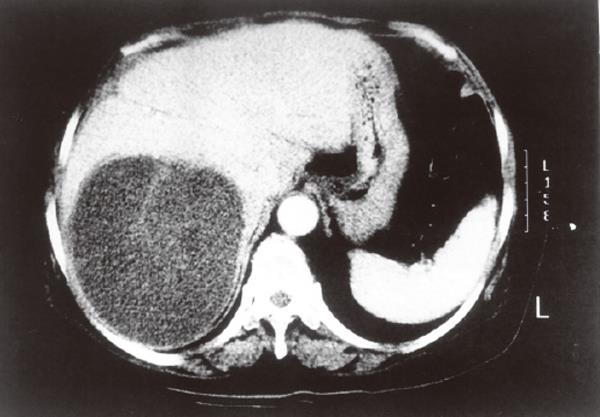
Figure 4 A big recurrent liver liposarcoma locating in the right lobe of the liver by CT.
Figure 5 A high grade of undifferentiated liposarcoma composed of spindle cells with marked nuclear pleomorphism (hematoxylin-eosin staining, × 400 ).
- Citation: Kuo LM, Chou HS, Chan KM, Yu MC, Lee WC. A case of huge primary liposarcoma in the liver. World J Gastroenterol 2006; 12(7): 1157-1159
- URL: https://www.wjgnet.com/1007-9327/full/v12/i7/1157.htm
- DOI: https://dx.doi.org/10.3748/wjg.v12.i7.1157


