Copyright
©2006 Baishideng Publishing Group Co.
World J Gastroenterol. Feb 21, 2006; 12(7): 1043-1048
Published online Feb 21, 2006. doi: 10.3748/wjg.v12.i7.1043
Published online Feb 21, 2006. doi: 10.3748/wjg.v12.i7.1043
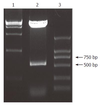
Figure 1 pGBKT7- HBcAg cut by EcoR I and Pst I on 0.
9% agarose/EtBr gel. Lanes 1 and 3: Marker; lane 2: HBcAg fragment cut by EcoR I and Pst I.
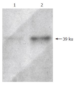
Figure 2 Western blotting shows the expression of HBcAg in yeast.
Lane 1 is negative control and lane 2 is HBV core protein.
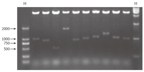
Figure 3 Screening colonies cut by BglII on 0.
9% agarose/EtBr gel
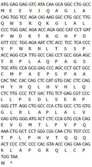
Figure 4 The nucleotide sequence of C1 gene and relevent amino acid sequences.
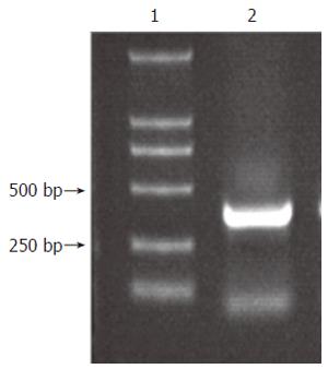
Figure 5 C1 DNA fragment amplified by RT-PCR.
Lane 1 is Marker and lane 2 is a 366 bp fragment of C1, amplified by RT-PCR.
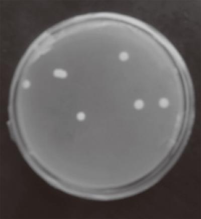
Figure 6 Positive clones interactive with the HBcAg protein grew on media lacking leucine, tryptophan, histidine and adenine.
- Citation: Lin SM, Cheng J, Lu YY, Zhang SL, Yang Q, Chen TY, Liu M, Wang L. Screening and identification of interacting proteins with hepatitis B virus core protein in leukocytes and cloning of new gene C1. World J Gastroenterol 2006; 12(7): 1043-1048
- URL: https://www.wjgnet.com/1007-9327/full/v12/i7/1043.htm
- DOI: https://dx.doi.org/10.3748/wjg.v12.i7.1043


