Copyright
©2006 Baishideng Publishing Group Co.
World J Gastroenterol. Feb 7, 2006; 12(5): 772-775
Published online Feb 7, 2006. doi: 10.3748/wjg.v12.i5.772
Published online Feb 7, 2006. doi: 10.3748/wjg.v12.i5.772
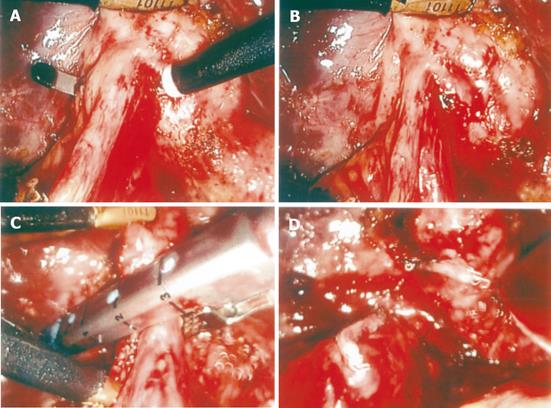
Figure 1 Procedures showing management of cholecystoenteric fistula.
A: Identification of cholecystocolic fistula; B: fistula is clearly exposed; C: fistula tract communicating with the transverse colon is resected using an Endo-GIA device; D: fistula is disconnected.
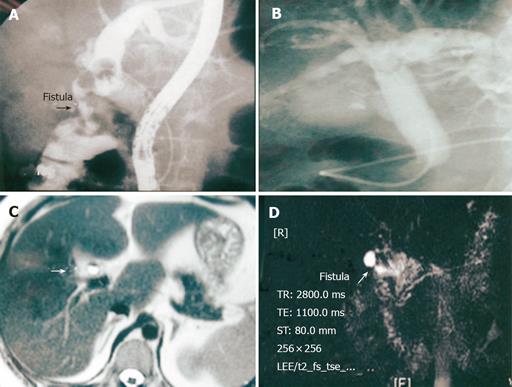
Figure 2 Detection of cholecystoenteric fistula radiologically.
A: ERCP showing appearance of pneumobilia with common bile duct stone and a fistula between collapsed gallbladder and transverse colon; B: ERCP showing common bile duct stone and pneumobilia; C: MRCP T2-weighed image showing pneumobilia and air-fluid level within the gallbladder; D: MRCP T2-weighed image revealing fistula between collapsed gallbladder and duodenum.
- Citation: Wang WK, Yeh CN, Jan YY. Successful laparoscopic management for cholecystoenteric fistula. World J Gastroenterol 2006; 12(5): 772-775
- URL: https://www.wjgnet.com/1007-9327/full/v12/i5/772.htm
- DOI: https://dx.doi.org/10.3748/wjg.v12.i5.772


