©2006 Baishideng Publishing Group Co.
World J Gastroenterol. Nov 28, 2006; 12(44): 7213-7215
Published online Nov 28, 2006. doi: 10.3748/wjg.v12.i44.7213
Published online Nov 28, 2006. doi: 10.3748/wjg.v12.i44.7213
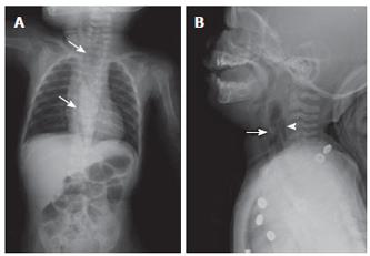
Figure 1 A: Extensive pneumomediastinum and air collections in retropharyngeal area (arrows), B: Irregular hyperdense lesion (arrow head) around cricopharyngeal area, and intra-esophageal opacification (arrow) was noted at C3 - C4 level.
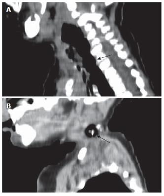
Figure 2 A: Esophageal rupture with retropharyngeal air collection (arrow), and B: Irregular foreign body impacted at upper esophagus (arrow).
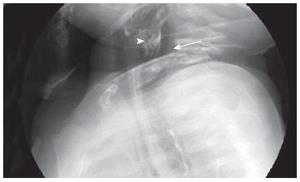
Figure 3 Much air collection (arrow) was in the retropharyngeal space and mediastinum, contrast leakage (arrow head) into the retropharyngeal space with opacification of a small fistulous tract at C3-4 level (cricopharyngeal level), suggestive of esophageal perforation.
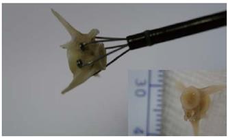
Figure 4 The fish vertebra with three sharp spines, which is about 1 cm x 1 cm in size, was removed by grasping forceps.
- Citation: Chang MY, Chang ML, Wu CT. Esophageal perforation caused by fish vertebra ingestion in a seven-month-old infant demanded surgical intervention: A case report. World J Gastroenterol 2006; 12(44): 7213-7215
- URL: https://www.wjgnet.com/1007-9327/full/v12/i44/7213.htm
- DOI: https://dx.doi.org/10.3748/wjg.v12.i44.7213


