Copyright
©2006 Baishideng Publishing Group Co.
World J Gastroenterol. Nov 7, 2006; 12(41): 6602-6607
Published online Nov 7, 2006. doi: 10.3748/wjg.v12.i41.6602
Published online Nov 7, 2006. doi: 10.3748/wjg.v12.i41.6602
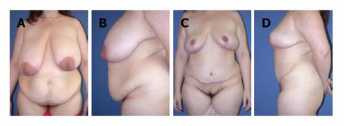
Figure 1 Typical frontal (A) and lateral (B) appearance of the anterior abdominal wall of a (non-weight loss) post-partum female.
In addition to excess skin and subcutaneous tissue, there is significant laxity of the anterior abdominal wall of this 50-year-old female; C and D: Post-operative appearance several months after standard abdominoplasty.
Figure 2 Typical frontal (A) and lateral (B) pre-operative appearance of a panniculectomy candidate; This 41-year-old patient is status post a 60-lb weight loss.
In addition to the unsightly appearance, the hanging pannus can be the source of pain and recurrent infection. Removal of the pannus (in this case with umbilical translocation) even without plication of the underlying abdominal wall resulted in significant improvement in contour and relief of symptoms (C and D). The patient also underwent a concurrent bilateral mastopexy.
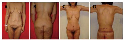
Figure 3 A 34-year-old female status post 120-lb weight loss.
Note the deflated, empty breasts and excess anterior abdominal skin and subcutaneous tissue anteriorly (A) and the excess skin and subcutaneous tissue posteriorly as well as the sagging buttocks (B); One month after circumferential abdominoplasty (and bilateral brachioplasty), there is significantly improved contour with re-creation of a feminine waistline (C); In addition, the buttocks have been lifted as a result of the circumferential excision (D). The patient is awaiting bilateral breast augmentation and bilateral medial thigh lift.
Figure 4 Pre-operative appearance of a 23-year-old post-partum female after 60 lb weight loss.
The breasts are deflated and ptotic (A and B). Status post bilateral breast augmentation (with saline prostheses) and right sided circumareolar mastopexy (C and D). Note the improved projection, especially in the upper pole. Additionally, the pre-operative asymmetry has been corrected.
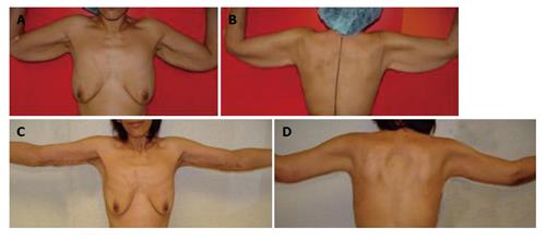
Figure 5 Typical appear-ance of upper extremity deformity after massive weight loss.
Note the “bat-wing” appearance of the proximal arm (A and B); One month after brachioplasty, there is significant improvement in contour (C and D). The incision is designed to keep the resultant scar on the medial surface of the upper extremity so that it is hidden when the patient stands at rest with her arms at her side.
- Citation: Spector JA, Levine SM, Karp NS. Surgical solutions to the problem of massive weight loss. World J Gastroenterol 2006; 12(41): 6602-6607
- URL: https://www.wjgnet.com/1007-9327/full/v12/i41/6602.htm
- DOI: https://dx.doi.org/10.3748/wjg.v12.i41.6602


