©2006 Baishideng Publishing Group Co.
World J Gastroenterol. Oct 21, 2006; 12(39): 6401-6404
Published online Oct 21, 2006. doi: 10.3748/wjg.v12.i39.6401
Published online Oct 21, 2006. doi: 10.3748/wjg.v12.i39.6401
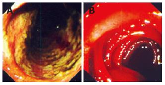
Figure 1 Endoscopic findings.
A: Extensive confluent necrotic ulcerations of the duodenal mucosa due to chemotherapy related S. stercoralis helminthic hyper-infection. B: Enteroscopic presentation of diffuse mucosal bleeding during anthelminthic therapy of S. stercoralis hyper-infection, aggravated potentially by toxic/inflammatory compounds from destroyed parasites.
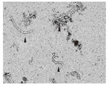
Figure 2 S.
stercoralis larvae in the stool.
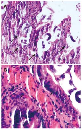
Figure 3 A: Intraluminal and intramucosal S.
stercoralis larvae in the duodenum during the exudative enteropathy hyper-infection phase (arrows). Scale: 200 μm. B: No intramural or peri-vascular inflammatory cell clusters are recognised during this early period of infection. Scale: 50 μm.
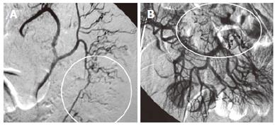
Figure 4 A: Segmental vasospasms (circled area).
B: “berry-type” micro-aneurysms (circled area) in the mesenteric superior and inferior arteries’ territory are both suggestive of active vasculitis during the anthelminthic therapy of S. stercoralis hyper-infection.
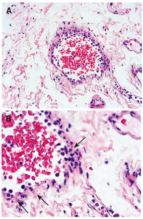
Figure 5 Perivascular granulocytic infiltration in the mucosa during the anthelminthic therapy of S.
stercoralis hyper-infection is also suggestive of active vasculitis. Scale: A: 200 μm; B: 50 μm.
- Citation: Csermely L, Jaafar H, Kristensen J, Castella A, Gorka W, Chebli AA, Trab F, Alizadeh H, Hunyady B. Strongyloides hyper-infection causing life-threatening gastrointestinal bleeding. World J Gastroenterol 2006; 12(39): 6401-6404
- URL: https://www.wjgnet.com/1007-9327/full/v12/i39/6401.htm
- DOI: https://dx.doi.org/10.3748/wjg.v12.i39.6401


