Copyright
©2006 Baishideng Publishing Group Co.
World J Gastroenterol. Oct 7, 2006; 12(37): 6008-6016
Published online Oct 7, 2006. doi: 10.3748/wjg.v12.i37.6008
Published online Oct 7, 2006. doi: 10.3748/wjg.v12.i37.6008
Figure 1 Modified manometry catheter for concurrent impedance-manometry procedure.
The 4 semiconductor solid-state pressure transducers (P1-P4) serve also as impedance electrodes and are placed at 6 cm distance each. The 8 impedance electrodes (4 mm length) are arranged between the pressure transducers at a distance of 16 mm. Together with the pressure transducers, they form 11 impedance segments, each 2 cm long (Ch1-Ch11). The solid-state pressure transducers are located exactly between the impedance channels 1-2, 4-5, 7-8, and 10-11, respectively. The first channels were located at the first part of the duodenum.
Figure 2 Classification of the peristaltic patterns according to Summers et al[16].
The peristaltic pattern can be classified to be (A) propagated single (double spike) wave contraction, (B) isolated (stationary) cluster of contractions, (C) propagated contraction with a clustered contraction, and (D) propagated cluster of contractions.
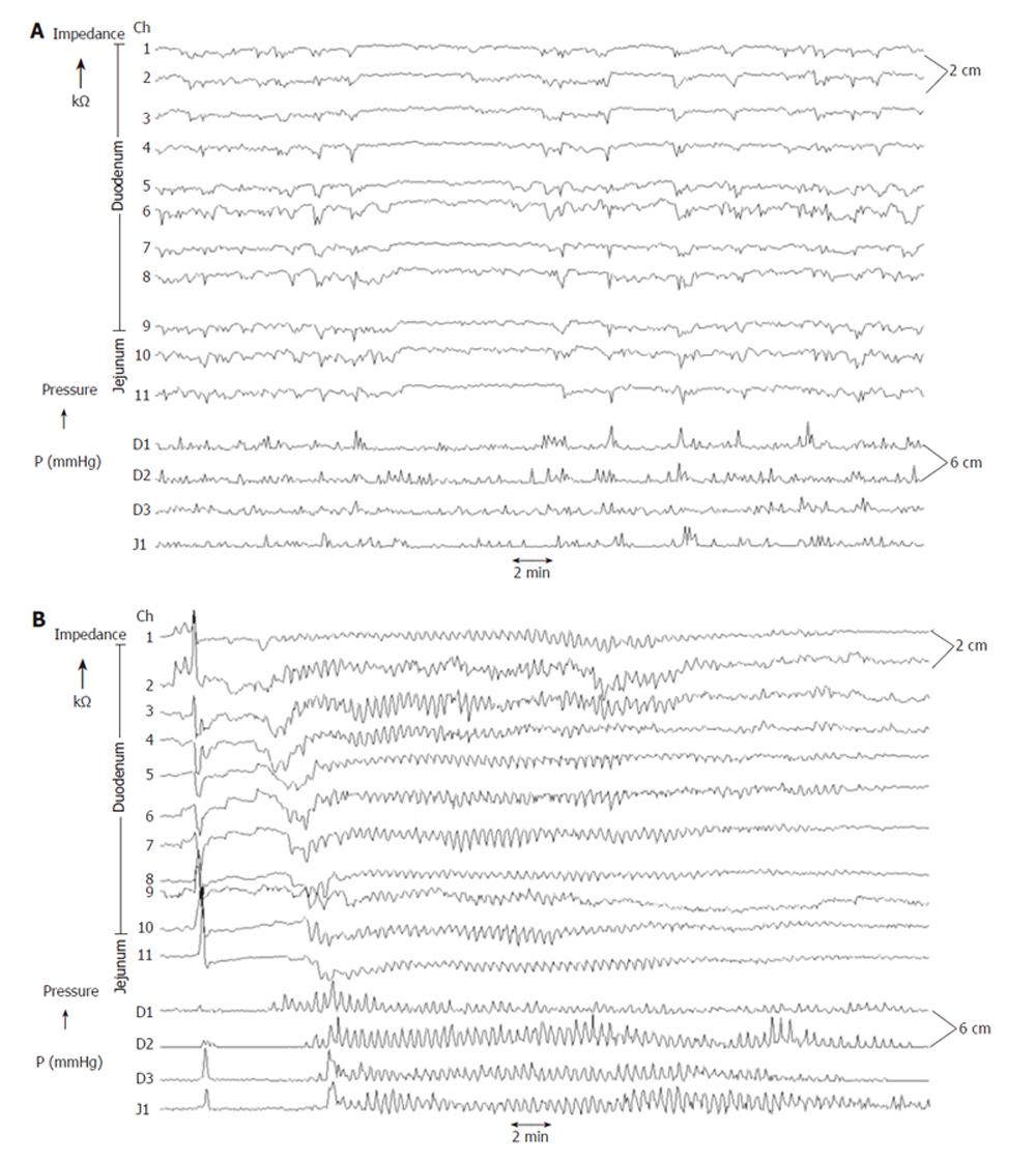
Figure 3 Concurrent Impedance Manometry (CIM) tracings.
Upper panel: During the interdigestive phase irregular chyme transport at the impedance channels and irregular motor activities at the pressure channels are observed. Lower panel: A phase 3 complex displays nearly identical features with frequent changes of both pressure and impedance.
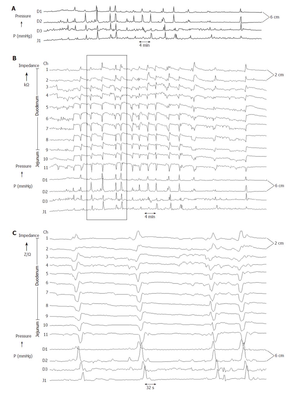
Figure 4 Concurrent Impedance Manometry (CIM) tracings after a test meal.
Upper panel: Low time scaled manometry tracings of the postprandial state. Middle panel: Low time scaled impedance manometry tracings of the same period as above showing several bolus transport events with associated peristaltic activities. Lower panel: High time scaled impedance manometry tracings of the box allowing identification and classification of bolus transport patterns as well as analysis of associated peristaltic patterns.
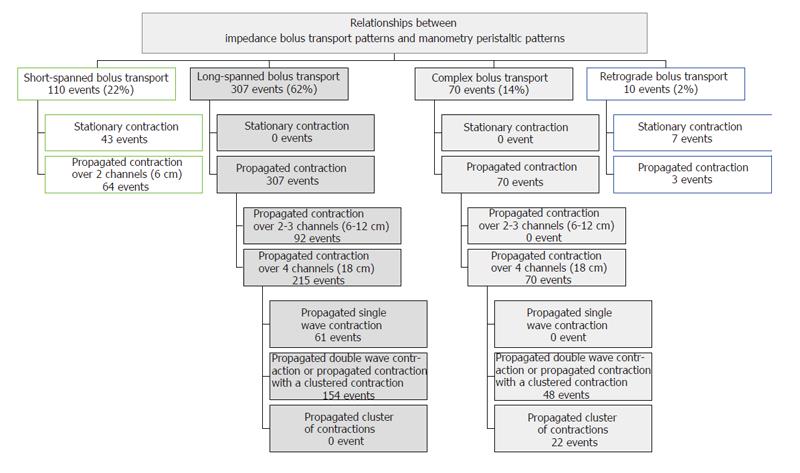
Figure 5 Distribution of bolus transport patterns and associated peristaltic patterns (see text for details).
Figure 6 Examples of simple bolus transports and associated peristalsis.
Upper panel: Short-spanned bolus transport with associated stationary contraction. Lower panel: Retrograde bolus transport with associated retrograde peristalsis.
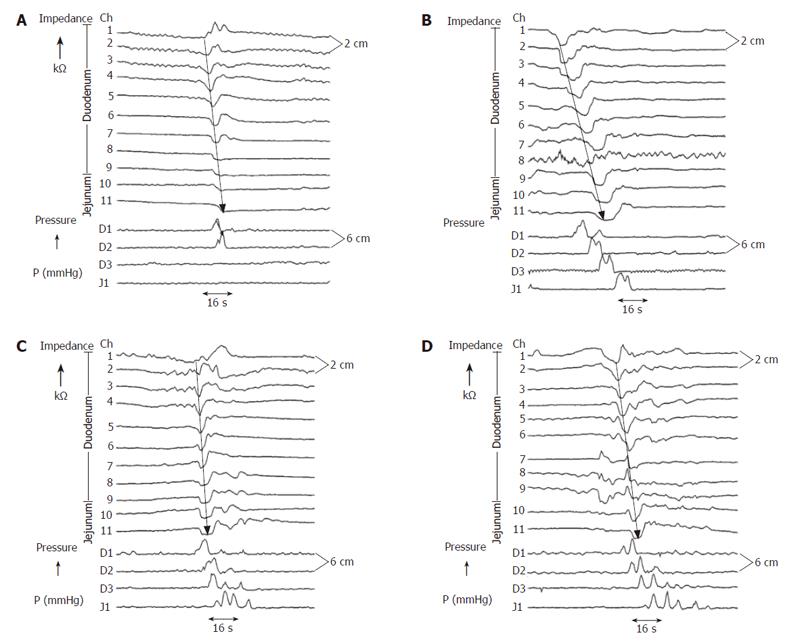
Figure 7 Examples of long-spanned bolus transports and associated peristalsis.
(A) A long-spanned bolus transport with the bolus front traversing the whole duodenum. The associated peristaltic sequence displayed a propagated single wave contraction from D1 to D2. This peristaltic pattern was classified to be simple (propagated single wave contraction). (B) A long-spanned bolus transport traversing the whole duodenum. The associated peristaltic sequence displayed a propagated contraction with a double wave contraction at channel D1 and double spike contractions at D2, D3 and J1. This peristaltic pattern was classified to be complex (propagated contraction with a double wave contraction). (C) A long-spanned bolus transport with the bolus front traversing the whole duodenum. The associated peristaltic sequence displayed single wave contractions at channel D1-D3 and a clustered contractions at J1. This peristaltic pattern was classified to be complex (propagated contraction with a clustered contraction). (D) A long-spanned bolus transport with the bolus front traversing the whole duodenum. The associated peristaltic sequence displayed double wave contractions at channel D1 and a clustered contractions at D2, D3 and J1. This peristaltic pattern was classified to be complex (propagated cluster of contractions).
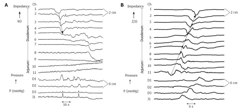
Figure 8 Examples of complex bolus transports and associated complex peristalsis.
(A) A complex bolus transport with two components propelling with different propulsion velocity as illustrating by the arrows. The associated peristaltic sequence is complex showing a long lasting double spike contraction at D1, a single wave contraction at D2, a clustered contraction at D3 and a multispike double wave contraction at J1. (B) A complex bolus transport comprising two boluses following each other very rapidly as illustrating by the anterograde arrows. The associated peristalsis showed two separate peristaltic sequences which seemed to be connected together by a interpolate contraction wave at D2. A retrograde bolus movement (retrograde arrow) seemed to be derive from a retrograde peristaltic sequence with a single contraction wave seen at J1.
- Citation: Nguyen HN, Winograd R, Domingues GRS, Lammert F. Postprandial transduodenal bolus transport is regulated by complex peristaltic sequence. World J Gastroenterol 2006; 12(37): 6008-6016
- URL: https://www.wjgnet.com/1007-9327/full/v12/i37/6008.htm
- DOI: https://dx.doi.org/10.3748/wjg.v12.i37.6008


