Copyright
©2006 Baishideng Publishing Group Co.
World J Gastroenterol. Sep 14, 2006; 12(34): 5565-5568
Published online Sep 14, 2006. doi: 10.3748/wjg.v12.i34.5565
Published online Sep 14, 2006. doi: 10.3748/wjg.v12.i34.5565
Figure 1 Barium swallow shows the presence of a stenotic trait in the middle thoracic esophagus.
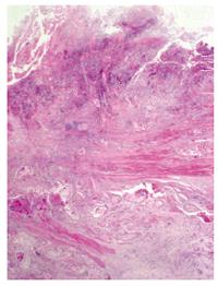
Figure 2 Esophageal histological sample (haematoxylin-eosin stain 20 x) shows the presence of squamous cell carcinoma with involvement of muscular tunic.
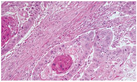
Figure 3 A squamous pearl at a higher magnification (haematoxylin-eosin stain 200 x).
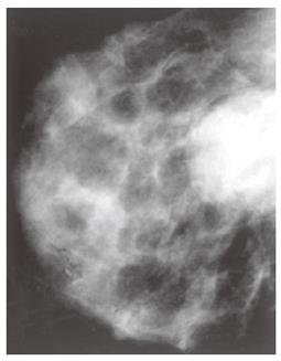
Figure 4 Mammography shows a well-defined nodule without calcification.
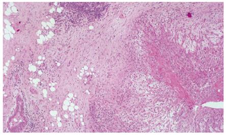
Figure 5 The right side of breast histological sample shows an infiltranting squamous carcinoma’s focus with peritumoral inflammation while the left side shows a normal mammary duct section (haematoxylin-eosin stain 40 x).
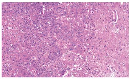
Figure 6 Squamous carcinoma infiltrating the cerebral parenchyma (haematoxylin-eosin stain 100 x).
- Citation: Santeufemia DA, Piredda G, Fadda GM, Rocca PC, Costantino S, Sanna G, Sarobba MG, Pinna MA, Putzu C, Farris A. Successful outcome after combined chemotherapeutic and surgical management in a case of esophageal cancer with breast and brain relapse. World J Gastroenterol 2006; 12(34): 5565-5568
- URL: https://www.wjgnet.com/1007-9327/full/v12/i34/5565.htm
- DOI: https://dx.doi.org/10.3748/wjg.v12.i34.5565


