©2006 Baishideng Publishing Group Co.
World J Gastroenterol. Sep 14, 2006; 12(34): 5440-5446
Published online Sep 14, 2006. doi: 10.3748/wjg.v12.i34.5440
Published online Sep 14, 2006. doi: 10.3748/wjg.v12.i34.5440
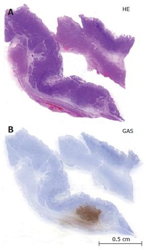
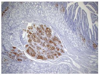
Figure 1 Overview of sporadic gastrinomas less than 3 mm in size within the submucosa stained with hematoxylin and eosin (A) and gastrin (B).
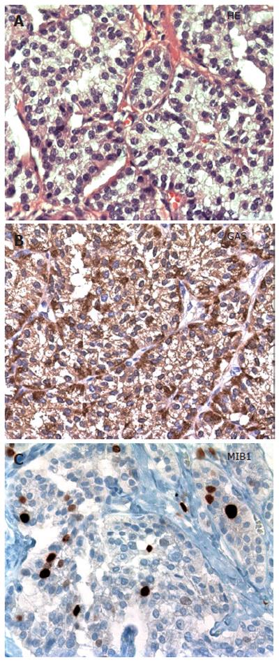
Figure 2 Morphology of sporadic duodenal gastrinoma stained with HE showing a trabecular and glandular growth pattern (A); strong immunoreactivity for gastrin (B), and expression of the nuclear proliferation antigen MIB1 in more than 2% of NET cells (C).
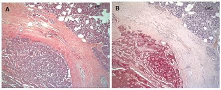
Figure 3 Overview of sporadic pancreatic gastrinoma surrounded by thickened collagen in the vicinity of normal pancreatic parenchyma stained with hematoxylin and eosin (A) and gastrin (B).
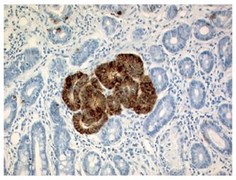
Figure 4 Circumscribed linear and nodular hyperplasia of gastrin cells within the Brunner’s glands in a patient with MEN1.
Figure 5 Tiny MEN1-associated duodenal gastrinoma within the submucosa revealing a diameter of less than 1 mm.
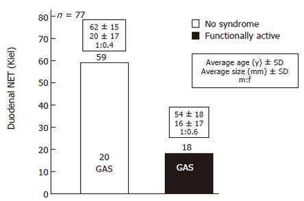
Figure 6 Duodenal NETs in the Kiel tumor archive.
Fifty-nine (76.6%) out of the 77 NETs were endocrinologically not active, 20 of them expressed gastrin. These were not associated with ZES. All the functionally active NETs (18; 23.4%) were immunohistochemically positive for gastrin and showed a ZES.
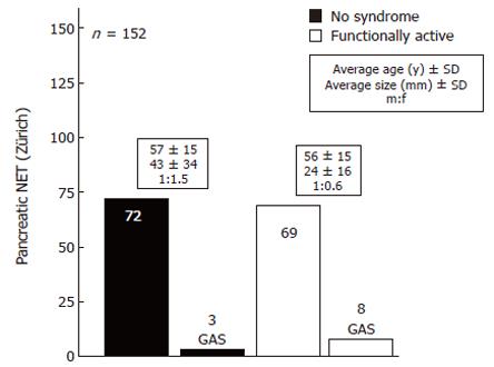
Figure 7 Pancreatic NETs in the Zürich tumor archives.
Seventy-five (49.3%) out of the 152 pancreatic NETs were endocrinologically not active, of which 3 expressed gastrin. These 3 gastrin-expressing NETs were not associated with ZES. Eight of the 77 functionally active NETs were immunohistochemically positive for gastrin and associated with ZES.
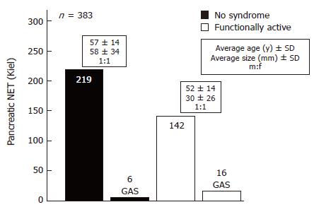
Figure 8 Pancreatic NETs in the Kiel tumor archives.
Two hundred and twenty-five (58.7%) out of the 383 pancreatic NETs were endocrinologically not active. These gastrin-expressing NET were not associated with ZES. Sixteen of the 158 functionally active NETs were immunohistochemically positive for gastrin and showed a ZES.
- Citation: Anlauf M, Garbrecht N, Henopp T, Schmitt A, Schlenger R, Raffel A, Krausch M, Gimm O, Eisenberger CF, Knoefel WT, Dralle H, Komminoth P, Heitz PU, Perren A, Klöppel G. Sporadic versus hereditary gastrinomas of the duodenum and pancreas: Distinct clinico-pathological and epidemiological features. World J Gastroenterol 2006; 12(34): 5440-5446
- URL: https://www.wjgnet.com/1007-9327/full/v12/i34/5440.htm
- DOI: https://dx.doi.org/10.3748/wjg.v12.i34.5440


