©2006 Baishideng Publishing Group Co.
World J Gastroenterol. Sep 7, 2006; 12(33): 5399-5400
Published online Sep 7, 2006. doi: 10.3748/wjg.v12.i33.5399
Published online Sep 7, 2006. doi: 10.3748/wjg.v12.i33.5399
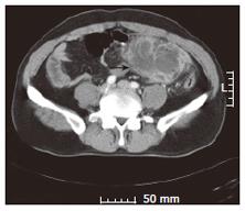
Figure 1 CT abdominal scan showing the presence of a solid fluid-containing tumor in the left lower quadrant (arrow).
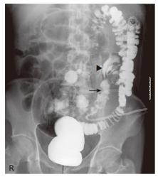
Figure 2 Radiograph using water-soluble contrast media showing the entire colon present in the left half of the abdomen, with the cecum in the left lower quadrant.
Contrast filling of the terminal ileum is shown (arrow). Irregular contour of the cecum (arrowhead) is shown with no contrast filling of the appendix. A pig-tail catheter is located at the area corresponding to the abscess pocket.
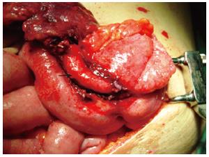
Figure 3 Intraoperative photography showing severe appendix infla-mmation (arrow) and the ileocecal region located in the left lower quadrant.
- Citation: Lee MR, Kim JH, Hwang Y, Kim YK. A left-sided periappendiceal abscess in an adult with intestinal malrotation. World J Gastroenterol 2006; 12(33): 5399-5400
- URL: https://www.wjgnet.com/1007-9327/full/v12/i33/5399.htm
- DOI: https://dx.doi.org/10.3748/wjg.v12.i33.5399


