©2006 Baishideng Publishing Group Co.
World J Gastroenterol. Aug 7, 2006; 12(29): 4673-4682
Published online Aug 7, 2006. doi: 10.3748/wjg.v12.i29.4673
Published online Aug 7, 2006. doi: 10.3748/wjg.v12.i29.4673
Figure 1 RT-PCR for the detection of HBx-RNA in HepG2 cells stably transfected HBx-expression constructs.
Total cellular RNA from clones obtained after stable transfection with expression vector pRCCMVX for wt-HBx (X1, X2, X3, X8, X10, and X18) or for inactive HBx (pRCCMVXM, XM1, XM2, XM6, XM12, XM14, and XM20) was amplified by RT-PCR with primers used for cloning the complete HBx-ORF.
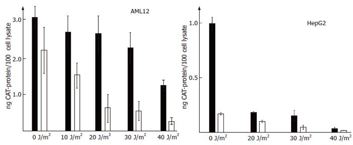
Figure 2 HBx activates the HBV-Enhancer I in vivo and can partially counteract a repression by p53 induced by UV irradiation.
CAT expression driven by Enhancer I of HBV is indicated as bars. AML12 (A) or HepG2 (B) derived cell lines expressing HBx (HepG2-X8 or AML12-HBx9) are indicated in black and the controls which did not express functional HBx (AML12 or HepG2-XM2) are shown in white bars. The cell lines were transfected with a CAT-expression vector driven by enhancer I of HBV in triplicate. Four hours after transfection, the cultures were irradiated with the indicated dose of UV. The lysis of the cells was done 18 h post irradiation.
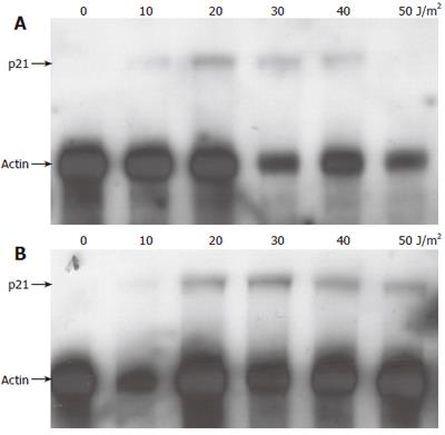
Figure 3 Induction of p53- and p21waf/cip/sdi-protein expression in HepG2-derived cell lines after irradiation with UV by immune blot.
The dose of UV used for irradiation is indicated. The cells were lysed 18 h after irradiation with UV.
Figure 4 Induction of p53- and p21waf/cip/sdi-protein expression in AML12-derived cell lines after irradiation with UV by immune blot.
The dose of UV used for irradiation is indicated. The cells were lysed 18 h after irradiation with UV.
Figure 5 Kinetics of induced p53- and p21waf/cip/sdi-protein expression after irradiation with UV by immune blot in AML12 cell lines.
The time in hours after irradiation with 40 J/m2 UV is indicated.
Figure 6 Detection of p21waf/cip/sdi-RNA by ribonuclease protection assay in the cell line HepG2-XM2 (A) and -HBx8 (B).
Total cellular RNA was isolated from cell cultures irradiated with the indicated dose of UV 18 h after irradiation. As internal control actin was detected by RPA.
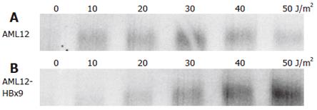
Figure 7 Detection of p21waf/cip/sdi-RNA by northern blot in the cell line AML12 (A) and the HBx-transformed clone AML12-HBx9 (B).
Total cellular RNA was isolated from cell cultures irradiated with the indicated dose of UV 18 h after irradiation.
- Citation: Fiedler N, Quant E, Fink L, Sun J, Schuster R, Gerlich WH, Schaefer S. Differential effects on apoptosis induction in hepatocyte lines by stable expression of hepatitis B virus X protein. World J Gastroenterol 2006; 12(29): 4673-4682
- URL: https://www.wjgnet.com/1007-9327/full/v12/i29/4673.htm
- DOI: https://dx.doi.org/10.3748/wjg.v12.i29.4673


