©2006 Baishideng Publishing Group Co.
World J Gastroenterol. Jul 21, 2006; 12(27): 4345-4351
Published online Jul 21, 2006. doi: 10.3748/wjg.v12.i27.4345
Published online Jul 21, 2006. doi: 10.3748/wjg.v12.i27.4345
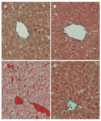
Figure 1 Histological appearance of rat liver tissue.
Masson X 270. A: Intact control; B: Control, zinc sulfate; The liver showing vacuolated hepatocytes (→) in the centrilobuler area with mild dilation of sinusoids (►) and hyperemia (*); C: Absolute ethanol; D: Zinc pre-treatment.
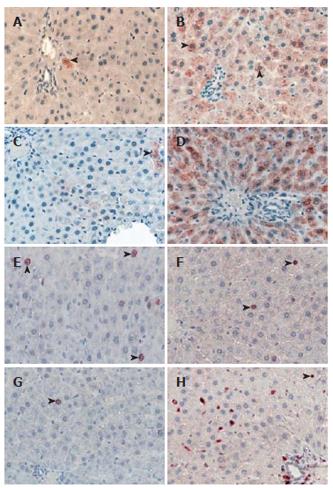
Figure 2 Immunreactive metallothionein-producing cells (►) in rat liver.
Streptavidin-Biotin-Peroxidase x 270. A: Intact control; B: Control, zinc sulfate; The reduced metallothionein expression (►); C: Ethanol; Note the increase of immunoreactivity of metallothionein-producing cells, D: Ethanol + zinc sulfate; E: Intact control, PCNA-positive hepatocytes (►); F: Control, zinc sulfate; G: Ethanol; The increase in immunoreactivity of PCNA-positive cells, H: Ethanol + zinc sulfate.
Figure 3 A: Metallothionein, aP = 0.
018 vs Ethanol; B: PCNA labeling indeces, bP = 0.007 vs Ethanol.
Figure 4 Ultrastructure of the hepatocytes of the rat.
TEM X5000. A: Control; B: Ethanol; C: Zinc sulfate + ethanol. An increase in smooth endoplasmic reticulum (SER), lipid vacuoles (L) and degeneration in mitochondria (M) of the hepatocyte of a rat given ethanol. These ultrastructural changes were improved by zinc pre-treatment.
- Citation: Bolkent S, Arda-Pirincci P, Bolkent S, Yanardag R, Tunali S, Yildirim S. Influence of zinc sulfate intake on acute ethanol-induced liver injury in rats. World J Gastroenterol 2006; 12(27): 4345-4351
- URL: https://www.wjgnet.com/1007-9327/full/v12/i27/4345.htm
- DOI: https://dx.doi.org/10.3748/wjg.v12.i27.4345


