Copyright
©2006 Baishideng Publishing Group Co.
World J Gastroenterol. Jun 14, 2006; 12(22): 3537-3545
Published online Jun 14, 2006. doi: 10.3748/wjg.v12.i22.3537
Published online Jun 14, 2006. doi: 10.3748/wjg.v12.i22.3537
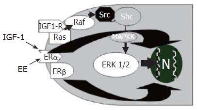
Figure 1 Additive effect of estrogens (EE) and insulin like growth factor-1 (IGF1) in the modulation of cholangiocyte proliferation.
EE and IGF1 play additive effects on cholangiocyte proliferation by acting at both receptor and post-receptor levels IGF1 and EE induce, in cholangiocytes, additive increase of phosphorylated ERK1/2.
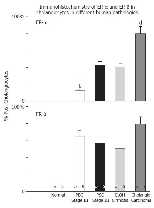
Figure 2 Immunohistochemistry of ER-α and ER-β in cholangiocytes in different human pathologies, bP < 0.
01 , dP < 0.01 vs other columns.
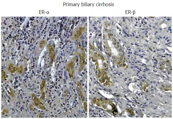
Figure 3 Immunohistochemistry for ER-α and ER-β.
Biopsies of human primary biliary cirrhosis showing an intense positivity for both ER-α and ER-β in the proliferating bile ducts. Orig. magn., x 20.
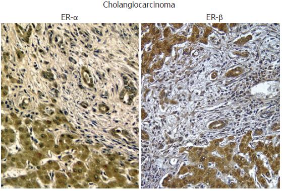
Figure 4 Immunohistochemistry for ER-α and ER-β.
Biopsies of human cholangiocarcinoma showing an intense positivity for both ER-α and ER-β. Orig. magn., x 20.
- Citation: Alvaro D, Mancino MG, Onori P, Franchitto A, Alpini G, Francis H, Glaser S, Gaudio E. Estrogens and the pathophysiology of the biliary tree. World J Gastroenterol 2006; 12(22): 3537-3545
- URL: https://www.wjgnet.com/1007-9327/full/v12/i22/3537.htm
- DOI: https://dx.doi.org/10.3748/wjg.v12.i22.3537


