©2006 Baishideng Publishing Group Co.
World J Gastroenterol. Jan 14, 2006; 12(2): 302-305
Published online Jan 14, 2006. doi: 10.3748/wjg.v12.i2.302
Published online Jan 14, 2006. doi: 10.3748/wjg.v12.i2.302
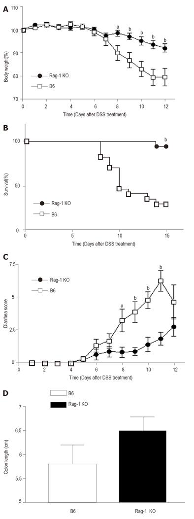
Figure 1 Rag-1 deficient mice showing resistance to develop of DSS-induced colitis.
Weight loss (A), mortality (B), the severity of diarrhea (C) and shortage of colon (D) after 1.5% DSS administration were analyzed. Each study group contained > 10 mice. Data shown represent mean values obtained from four independent experiments (aP<0.05; bP<0.01).
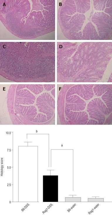
Figure 2 Hematoxylin and eosin (HE) staining of colons obtained from B6 and Rag-1 KO mice treated with or without DSS.
A, C: B6 mice treated with DSS (X100 and 160); B, D: Rag-1 KO mice treated with DSS (X100 and 160); E: B6 mice treated with water (X100); F: Rag-1 KO mice treated with water (X100); G: Quantitative histopathologic assessment of DSS-induced colitis activity showing a significant suppression in Rag-1 KO mice compared to the B6 control mice. Samples were collected from B6 control and Rag-1 KO mice treated with 1.5% DSS (open and solid bar, respectively), or water (stripped bars). Data are expressed as mean ± SE and represent > 10 mice per group (aP<0.05; bP<0.01).
- Citation: Kim TW, Seo JN, Suh YH, Park HJ, Kim JH, Kim JY, Oh KI. Involvement of lymphocytes in dextran sulfate sodium-induced experimental colitis. World J Gastroenterol 2006; 12(2): 302-305
- URL: https://www.wjgnet.com/1007-9327/full/v12/i2/302.htm
- DOI: https://dx.doi.org/10.3748/wjg.v12.i2.302


