Copyright
©2006 Baishideng Publishing Group Co.
World J Gastroenterol. Jan 14, 2006; 12(2): 259-264
Published online Jan 14, 2006. doi: 10.3748/wjg.v12.i2.259
Published online Jan 14, 2006. doi: 10.3748/wjg.v12.i2.259
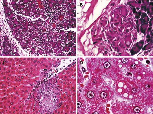
Figure 1 Histopathological changes in pancreatic and hepatic specimens.
A: Obvious acinar cell degeneration, edema and inflammation in caerulein group. B: Acinar cell degeneration and fat necrosis in caerulein group. C: Hepatocyte necrosis, vascular congestion, sinusoidal dilatation and inflammatory infiltration in caerulein group. D: Prominent intracellular vacuolization in hepatic specimens from caerulein group.
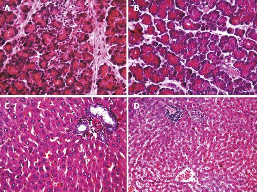
Figure 2 Histopathological evidence of pancreatic and hepatic damage.
A: Normal histological appearance except for minimal infiltration in caerulein + melatonin group. B: Normal histological appearance except for minimal edema in caerulein + L(+)-ascorbic acid + N-acetyl cysteine group. C:Normal histological appearance in caerulein + melatonin group. D: Two small areas of necrosis and cell infiltration in caerulein + L(+) - ascorbic acid + N - acetyl cysteine group.
-
Citation: Eşrefoğlu M, Gül M, Ateş B, Batçıoğlu K, Selimoğlu MA. Antioxidative effect of melatonin, ascorbic acid and
N -acetylcysteine on caerulein-induced pancreatitis and associated liver injury in rats. World J Gastroenterol 2006; 12(2): 259-264 - URL: https://www.wjgnet.com/1007-9327/full/v12/i2/259.htm
- DOI: https://dx.doi.org/10.3748/wjg.v12.i2.259


