Copyright
©2006 Baishideng Publishing Group Co.
World J Gastroenterol. May 14, 2006; 12(18): 2945-2948
Published online May 14, 2006. doi: 10.3748/wjg.v12.i18.2945
Published online May 14, 2006. doi: 10.3748/wjg.v12.i18.2945
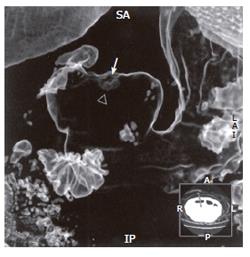
Figure 1 The ulcero carcinoma of stomach (on the anterosuperior wall of gastric antrum): the anterior-view of stomach on the VR image shows clearly the hollow lesion (↑) and the concomitant prominent lesion (△), with gas filling the stomach.
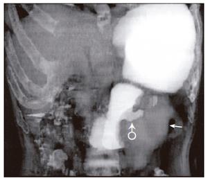
Figure 2 The ulcero carcinoma of stomach (on the greater curvature of stomach): the anterior-view of stomach on VR image shows the prominent lesion (↑) and the subsequent hollow lesion (♂), with 2% - 3% Omnipaque 300 filling the stomach.
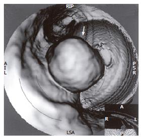
Figure 3 The leiomyoma of stomach (on the greater curvature of stomach): the axial-view of gastric cavity on VE image shows the prominent lesion (↑), with gas filling the stomach.
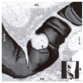
Figure 4 The same patient of Figure 3: the gastric coronal plane on MPR image shows the prominent lesion (↑) and the subsequent hollow lesion (♂).
Figure 5 The polypus of colon: the anterior-view of colon on SSD image, by using the cutting technique, shows the prominent lesion (↑), with air filling the large intestine.
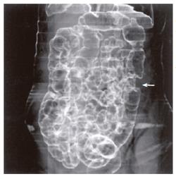
Figure 6 The carcinoma of descending colon: the anterior-view of large intestine on VR image shows the stenosis of intestinal cavity (↑), with air filling the large intestine.
- Citation: Duan SY, Zhang DT, Lin QC, Wu YH. Clinical value of CT three-dimensional imaging in diagnosing gastrointestinal tract diseases. World J Gastroenterol 2006; 12(18): 2945-2948
- URL: https://www.wjgnet.com/1007-9327/full/v12/i18/2945.htm
- DOI: https://dx.doi.org/10.3748/wjg.v12.i18.2945


