©2006 Baishideng Publishing Group Co.
World J Gastroenterol. Apr 7, 2006; 12(13): 2139-2142
Published online Apr 7, 2006. doi: 10.3748/wjg.v12.i13.2139
Published online Apr 7, 2006. doi: 10.3748/wjg.v12.i13.2139
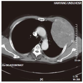
Figure 1 Chest helical CT.
A mass on the anterolateral aspect of the left chest wall. The inner part of the mass is heterogeneously attenuated and consistent with mottled calcification. There is no evidence of metastasis in the mediastinum or the lung parenchyma.
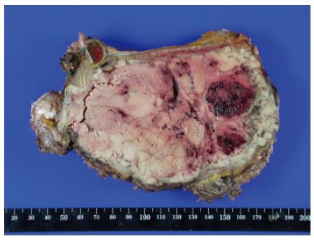
Figure 2 Gross findings of the chest wall tumor.
The cut surface shows an encapsulated, yellowish-white solid mass with areas of hemorrhage and necrosis.
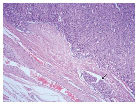
Figure 3 Microscopic findings of the chest wall tumor.
The tumor displays typical features of well-differentiated, trabecular liver cell carcinoma with intravascular tumor emboli (arrow) (H&E stain, ×200).
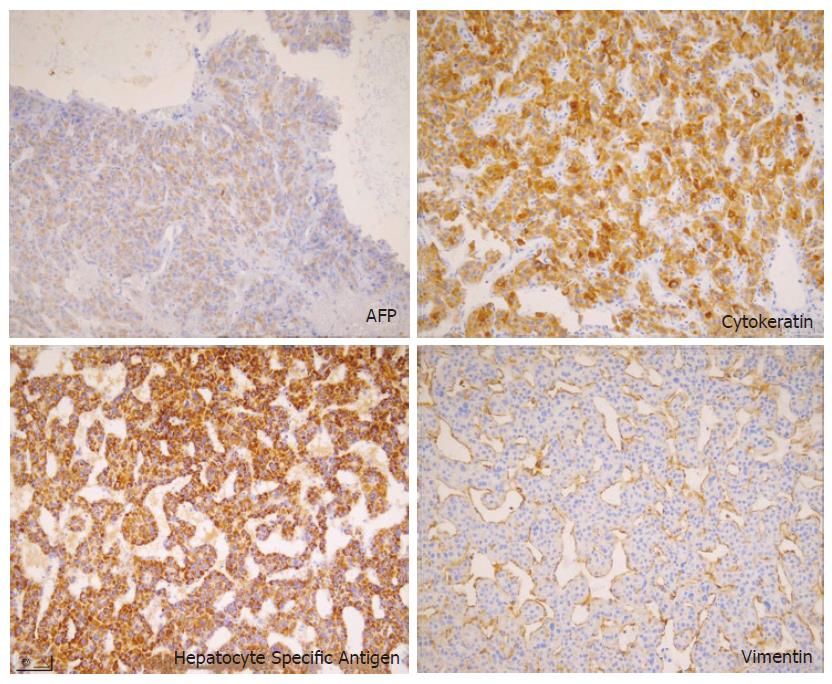
Figure 4 Microscopic findings of the chest wall tumor.
The tumor cells are positive for AFP, cytokeratin, and hepatocyte specific antigens. This section shows positive staining for hepatocyte-specific antigens (IHC, ×400).
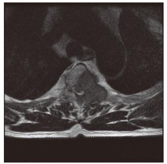
Figure 5 Thoracic spine MRI.
The metastatic tumor of the third vertebra compressing the spinal cord (T2 weighted image).
- Citation: Hyun YS, Choi HS, Bae JH, Jun DW, Lee HL, Lee OY, Yoon BC, Lee MH, Lee DH, Kee CS, Kang JH, Park MH. Chest wall metastasis from unknown primary site of hepatocellular carcinoma. World J Gastroenterol 2006; 12(13): 2139-2142
- URL: https://www.wjgnet.com/1007-9327/full/v12/i13/2139.htm
- DOI: https://dx.doi.org/10.3748/wjg.v12.i13.2139


