Copyright
©2006 Baishideng Publishing Group Co.
World J Gastroenterol. Mar 14, 2006; 12(10): 1649-1651
Published online Mar 14, 2006. doi: 10.3748/wjg.v12.i10.1649
Published online Mar 14, 2006. doi: 10.3748/wjg.v12.i10.1649
Figure 1 Specimen of the pancreaticoduodenectomy with negative margin.
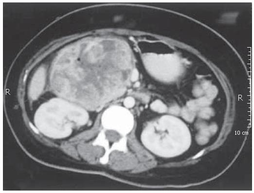
Figure 2 Computed tomography showing a 10 cm x 15 cm oval, mildly hypoechoic tumor with smooth margins.
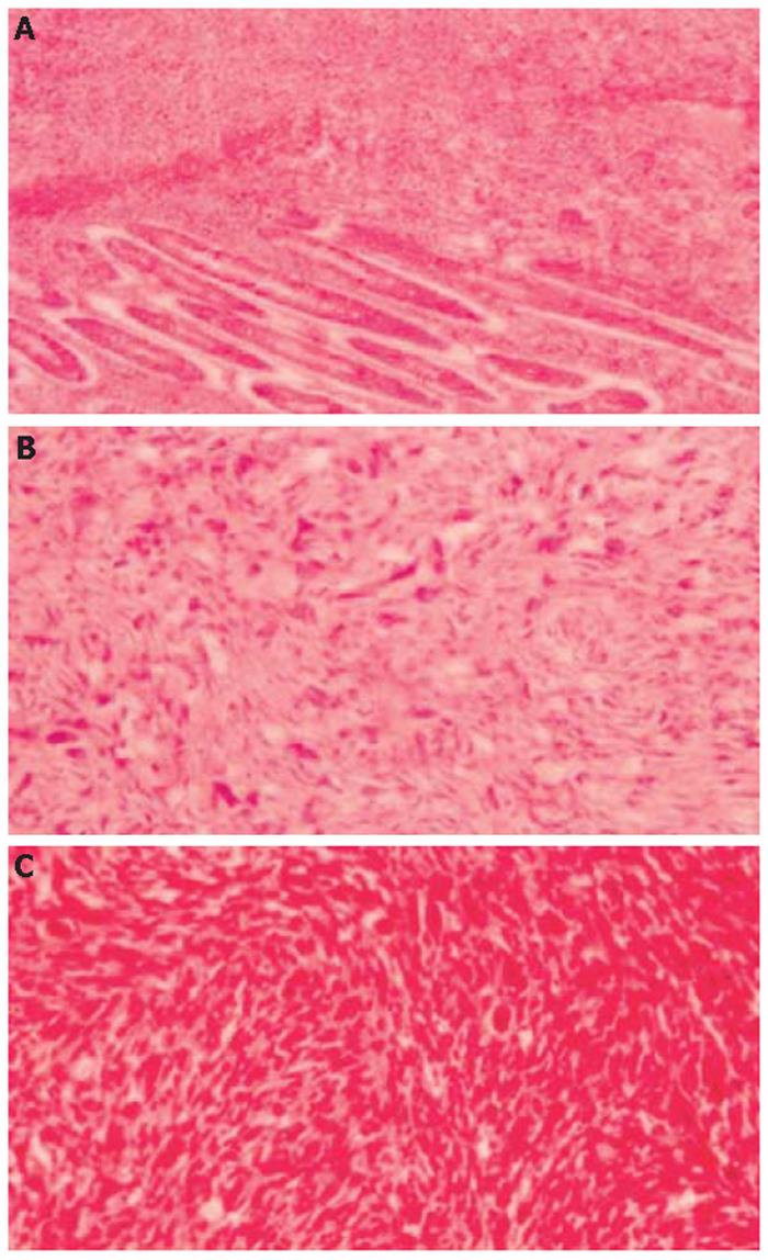
Figure 3 Neoplastic cells showing invasion of duodenal mucosa and submucosa (A) (H&E x 100); duodenal tumor consisted of haphazardly arranged pleomorphic spindle cells (B) (H&E x 200); breast tumor showing the similar histopathological findings with the duodenal tumor (C) (H&E x 200).
- Citation: Asoglu O, Karanlik H, Barbaros U, Yanar H, Kapran Y, Kecer M, Parlak M. Malignant phyllode tumor metastatic to the duodenum. World J Gastroenterol 2006; 12(10): 1649-1651
- URL: https://www.wjgnet.com/1007-9327/full/v12/i10/1649.htm
- DOI: https://dx.doi.org/10.3748/wjg.v12.i10.1649


