©2006 Baishideng Publishing Group Co.
World J Gastroenterol. Jan 7, 2006; 12(1): 146-149
Published online Jan 7, 2006. doi: 10.3748/wjg.v12.i1.146
Published online Jan 7, 2006. doi: 10.3748/wjg.v12.i1.146
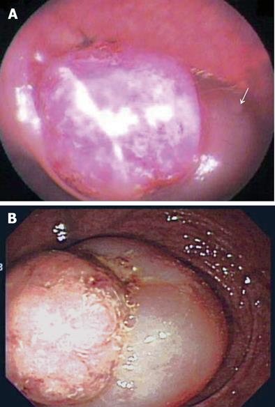
Figure 1 Endoscopic findings.
A: Flexible sigmoidoscopy at a local clinic showing the mass lesion 15 cm from the anal verge, and the partial downward displacement of involved bowel (arrow); B: Colonoscopy at our clinic showing the invaginated bowel with a round mass lesion about 3 cm from the anal verge.
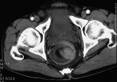
Figure 2 Contrast enhanced CT scan of pelvis showing a homogenously well enhancing mass at the intussuscepted bowel tip.
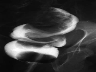
Figure 3 Gastrograffin study showing a large smooth-invaginated mass intussuscepting into the rectum.
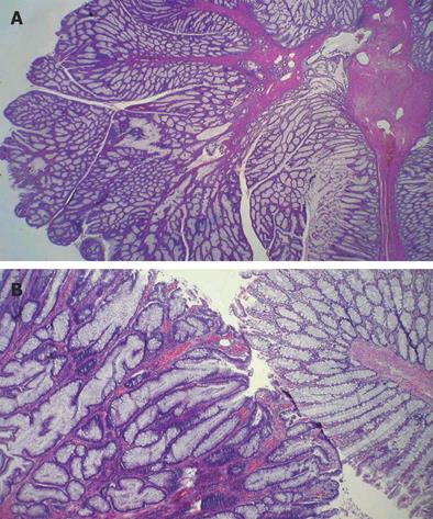
Figure 4 Hematoxylin and eosin staining of the lesions.
A: The polypoid mass of the colon showing branching papillary projections composed of proliferated glands and thin fibrous stalks (× 10); B: The proliferated glands of the polyp (left) lined by single or pseudostratified hyperchromatic columnar cells as compared with normal colonic mucosa (right), but without any malignant changes (× 40).
- Citation: Park KJ, Choi HJ, Kim SH, Han SY, Hong SH, Cho JH, Kim HH. Sigmoidorectal intussusception of adenoma of sigmoid colon treated by laparoscopic anterior resection after sponge-on-the-stick-assisted manual reduction. World J Gastroenterol 2006; 12(1): 146-149
- URL: https://www.wjgnet.com/1007-9327/full/v12/i1/146.htm
- DOI: https://dx.doi.org/10.3748/wjg.v12.i1.146


