Copyright
©2005 Baishideng Publishing Group Inc.
World J Gastroenterol. Nov 7, 2005; 11(41): 6489-6494
Published online Nov 7, 2005. doi: 10.3748/wjg.v11.i41.6489
Published online Nov 7, 2005. doi: 10.3748/wjg.v11.i41.6489
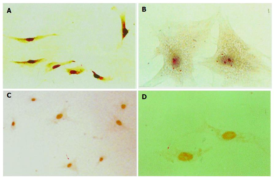
Figure 1 Immunocytochemistry and ISH detection of AT1 in hPSCs at protein and mRNA levels.
Immunostaining positive particles were located in plasm, A (magnification ×100); B (magnification ×400). ISH positive signals were located in nuclei C (magnification ×100); D (magnification ×400).
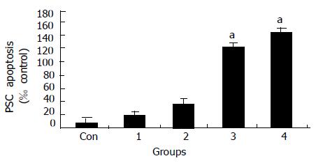
Figure 2 Dose-dependent induction of apoptosis by Losartan at different concentrations.
(Con, 1 = 10-9, 2 = 10-7 , 3 = 10-5, 4 = 10-3 mol/L; aP < 0.05 vs different from all groups ).
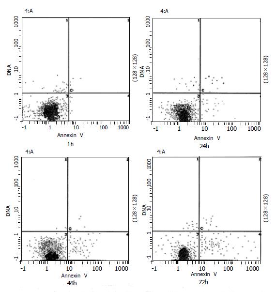
Figure 3 Time-course of Losartan-induced apoptosis.
hPSCs in 0.1% FCS were exposed to 10-5 mol/L Losartan, and was assessd at 24, 48,72 h. Results indicate that Losartan induces apoptosis in a time-dependent manner.
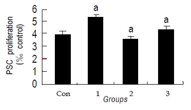
Figure 4 Effects of Losartan on hPSCs proliferation assessed with BrdU incorporation.
Losartan itself did not affect cell proliferation compared to control group. (aP>0.05 different from all the groups).
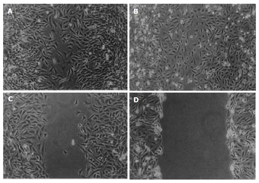
Figure 5 AngII accelerates in vitro wound healing confluent, culture-activated PSCs were serum deprived for 48 h.
A wound was produced in the monolayer with a pipette tip, and the cells were exposed to (A) AngII (10-8 mol/L), (B) 10% fetal bovine serum, (C) AngII + Losartan, (D) serum-free medium along 24 h later, the cells were photographed.
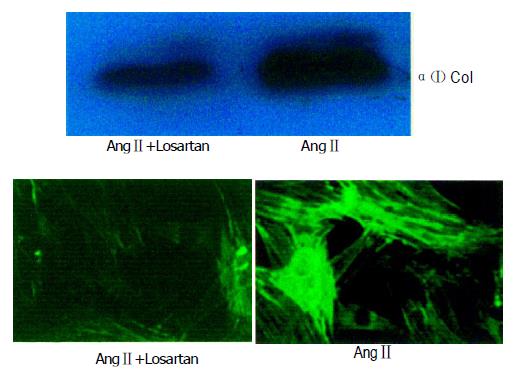
Figure 6 Losartan down-regulated collagen I protein expression by western blot and immunofluoresence.
hPSCs were incubated with 10-6 mol/L Losartan. Samples were exposed to certain concentration of Losartan+AngII and AngII for 24 h.
- Citation: Liu WB, Wang XP, Wu K, Zhang RL. Effects of angiotensin II receptor antagonist, Losartan on the apoptosis, proliferation and migration of the human pancreatic stellate cells. World J Gastroenterol 2005; 11(41): 6489-6494
- URL: https://www.wjgnet.com/1007-9327/full/v11/i41/6489.htm
- DOI: https://dx.doi.org/10.3748/wjg.v11.i41.6489


