Copyright
©2005 Baishideng Publishing Group Inc.
World J Gastroenterol. Oct 28, 2005; 11(40): 6381-6384
Published online Oct 28, 2005. doi: 10.3748/wjg.v11.i40.6381
Published online Oct 28, 2005. doi: 10.3748/wjg.v11.i40.6381
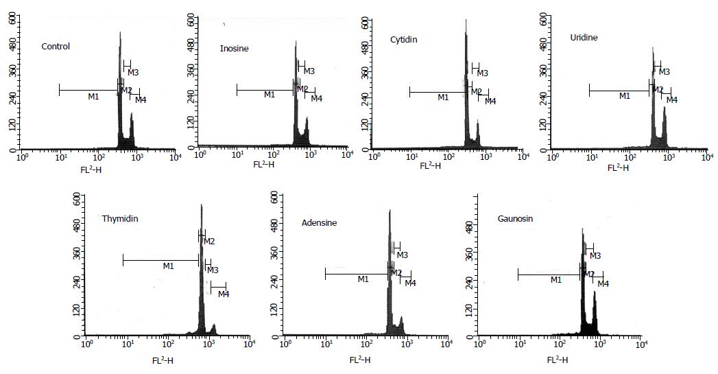
Figure 1 Effects of nucleosides on the cell cycle progression of HepG2 cells.
HepG2 cells untreated (control) or treated with nucleosides were stained with propidium iodide and analyzed using a flow cytometer. The contents of inosine, cytidine, uridine, and thymidine in mediums were 30 mmol/L, respectively. And, the contents of adenosine and guanosine were 0.5 mmol/L. This experiment was repeated thrice and obtained similar results were obtained. Figure 1 was the example from all results in each group. M1, M2, M3, and M4 represented Sub G1, G0/G1, S, and G2/M phases, respectively (A-G).
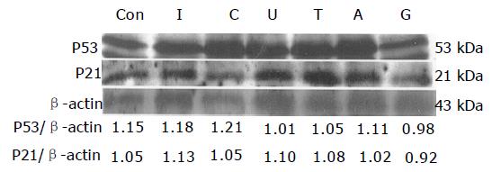
Figure 2 Effects of nucleosides on the p53 and p21 protein expressions in HepG2 cells.
One hundred μg of cytosolic proteins were separated by SDS-PAGE, and P53, P21, and β-actin protein were detected, respectively. This experiment was repeated thrice and similar results obtained.
- Citation: Yang SC, Chiu CL, Huang CC, Chen JR. Apoptosis induced by nucleosides in the human hepatoma HepG2. World J Gastroenterol 2005; 11(40): 6381-6384
- URL: https://www.wjgnet.com/1007-9327/full/v11/i40/6381.htm
- DOI: https://dx.doi.org/10.3748/wjg.v11.i40.6381


