Copyright
©2005 Baishideng Publishing Group Inc.
World J Gastroenterol. Oct 28, 2005; 11(40): 6348-6353
Published online Oct 28, 2005. doi: 10.3748/wjg.v11.i40.6348
Published online Oct 28, 2005. doi: 10.3748/wjg.v11.i40.6348
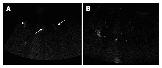
Figure 1 Pre-contrast image in a 32-year-old normal control (A) and contrast-enhanced US image in a 64-year-old cirrhotic patient (B).
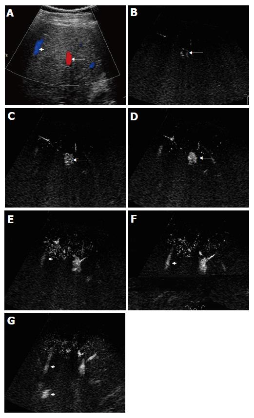
Figure 2 Contrast-enhanced US images in a 32-year-old normal control.
A: Before contrast, color Doppler US was performed to show the location and course of the portal vein (arrow) and hepatic vein (arrowhead); B: In the early arterial phase, the portal vein was not enhanced, while the parallel running artery was displayed by microbubble enhancement (arrow); C: Twenty-eight seconds after injection of Levovist, bright dots of microbubbles began to fill in the portal vein (arrow); D: Four seconds later, the portal vein was obviously enhanced by microbubbles (arrow); E: Before microbubbles arrived, the hepatic vein (arrowhead) was mildly echogenic; F and G: Thirty-seven seconds after injection of contrast medium, the hepatic vein (arrowhead) became more echogenic (F) than it was on E, indicating enhancement by microbubbles, and 4 s later the proximal part of hepatic vein was also clearly seen (arrowheads) (G).
Figure 3 Contrast-enhanced US images in normal controls.
A: In the early arterial phase, only the peripheral arteries were displayed, they were thin and regular; B and C: In the vascular phase of contrast-enhanced US, the peripheral portal vein (B) and hepatic vein (C) were portrayed as regular in caliber, course, and ramification, tapering smoothly as they run to the liver surface.
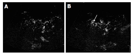
Figure 4 Contrast-enhanced US images in cirrhotic patients.
A and B: On contrast-enhanced US images, the peripheral vessels had poor delineation and were irregular (A). There was no tapering of the vessels. Small branches of the vessels were rarely seen (A), while abnormal ring-like structure (B, arrow) was displayed.
Figure 5 Contrast-enhanced US images in a 64-year-old cirrhotic patient.
A: In the early arterial phase 14 s after Levovist injection, a “Y”-shaped distal portal venous branch was enhanced by microbubbles (arrow), while the proximal portal vein and other portal venous branches at the same level were not seen; B and C: An adjacent hepatic venous branch was enhanced by contrast medium at 16 s after injection of Levovist (arrows).
- Citation: Zheng RQ, Zhang B, Kudo M, Sakaguchi Y. Hemodynamic and morphologic changes of peripheral hepatic vasculature in cirrhotic liver disease: A preliminary study using contrast-enhanced coded phase inversion harmonic ultrasonography. World J Gastroenterol 2005; 11(40): 6348-6353
- URL: https://www.wjgnet.com/1007-9327/full/v11/i40/6348.htm
- DOI: https://dx.doi.org/10.3748/wjg.v11.i40.6348


