Copyright
©The Author(s) 2005.
World J Gastroenterol. Oct 21, 2005; 11(39): 6125-6129
Published online Oct 21, 2005. doi: 10.3748/wjg.v11.i39.6125
Published online Oct 21, 2005. doi: 10.3748/wjg.v11.i39.6125
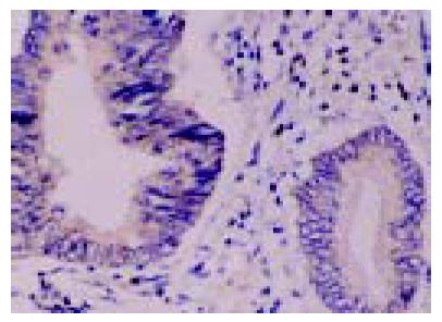
Figure 1 Immunohistochemical staining of Fas and FasL expression in colon of FasL in colon cancer cells; and C: high expression of FasL in lymph node cancer cells.
A: Low expression of Fas in colon cancer cells; B: high expression metastases from colon cancer cells.
Figure 2 Localization of FasL mRNA by in situ hybridization.
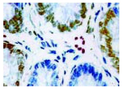
Figure 3 Detection of TILs.
They were displayed by double staining (CD45 staining and TUNEL).
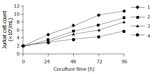
Figure 4 Jurkat cells growth curve after being co-cultured with SW480.
1: Control group; 2: 0.5×10⁵/mL SW480 planting group; 3: 1×10⁵/mL SW480 planting group; and 4: 2×10⁵/mL SW480 planting group.
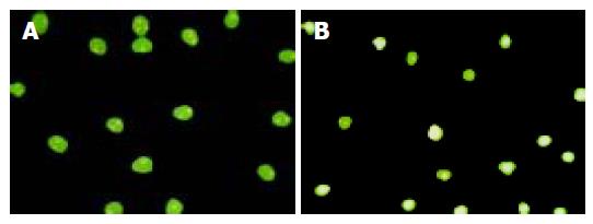
Figure 5 Detection of apoptotic Jurkat cells by fluorescence microscopy.
A: Negative control of Jurkat cells; B: apoptotic Jurkat cells after being co-cultured with SW480 cells.
- Citation: Zhu Q, Liu JY, Xu HW, Yang CM, Zhang AZ, Cui Y, Wang HB. Mechanism of counterattack of colorectal cancer cell by Fas/Fas ligand system. World J Gastroenterol 2005; 11(39): 6125-6129
- URL: https://www.wjgnet.com/1007-9327/full/v11/i39/6125.htm
- DOI: https://dx.doi.org/10.3748/wjg.v11.i39.6125


