Copyright
©2005 Baishideng Publishing Group Inc.
World J Gastroenterol. Sep 21, 2005; 11(35): 5492-5497
Published online Sep 21, 2005. doi: 10.3748/wjg.v11.i35.5492
Published online Sep 21, 2005. doi: 10.3748/wjg.v11.i35.5492
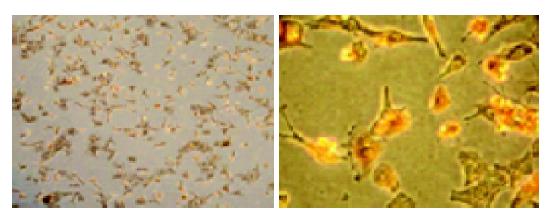
Figure 1 Cardiac myocytes identified by immunocytochemical assay.
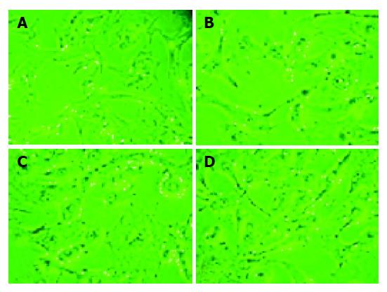
Figure 2 Cellular morphological change of cardiomyocytes after exposure to different concentrations of hydrogen peroxide for 4 h.
A: Normal control group (magnification ×200); B: cardiomyocytes exposure to 1.25 mmol/L H2O2 (magnification ×200); C: cardiomyocytes exposure to 2.5 mmol/L H2O2 (magnification ×200); D: cardiomyocytes exposure to 5 mmol/L H2O2 (magnification ×200).
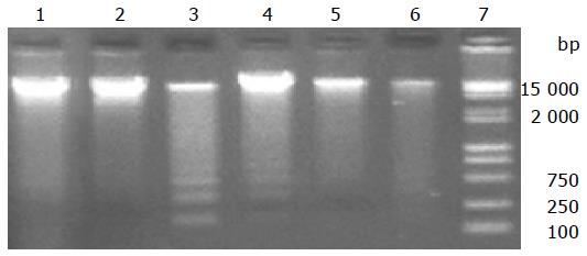
Figure 3 Electrophoresis analysis of DNA extracted from cultured myocytes.
Lane 1: normal control group; lane 2: 0.625 mmol/L H2O2 for 3 h; lane 3: 1.25 mmol/L H2O2 for 3 h; lane 4: 2.5 mmol/L H2O2 for 3 h; lane 5: 5 mmol/L H2O2 for 3 h; lane 6: 10 mmol/L H2O2 for 3 h; lane 7: DNA marker.
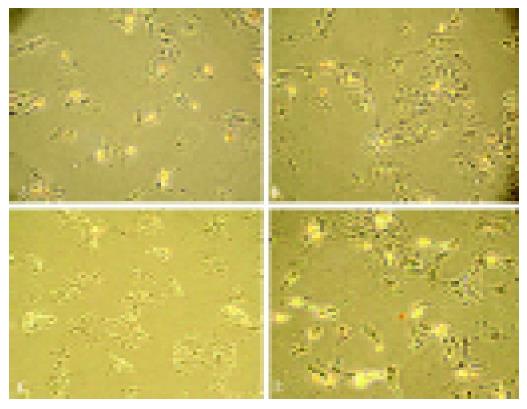
Figure 4 Anti-apoptosis effect of nm-haFGF on cardiomyocytes injured by H2O2.
- Citation: Lin ZF, Li XK, Lin Y, Wu F, Liang LM, Fu XB. Protective effects of non-mitogenic human acidic fibroblast growth factor on hydrogen peroxide-induced damage to cardiomyocytes in vitro. World J Gastroenterol 2005; 11(35): 5492-5497
- URL: https://www.wjgnet.com/1007-9327/full/v11/i35/5492.htm
- DOI: https://dx.doi.org/10.3748/wjg.v11.i35.5492


