Copyright
©The Author(s) 2005.
World J Gastroenterol. Sep 14, 2005; 11(34): 5303-5308
Published online Sep 14, 2005. doi: 10.3748/wjg.v11.i34.5303
Published online Sep 14, 2005. doi: 10.3748/wjg.v11.i34.5303
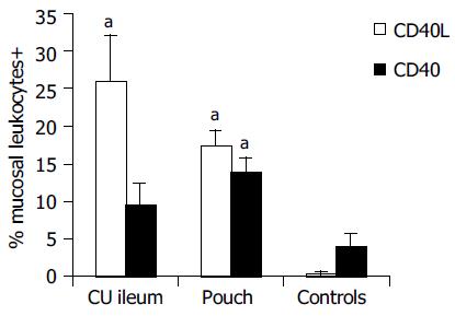
Figure 1 Mean percentages with standard error of CD40L+ and CD40+ leukocytes in the mucosa of UC ileum, ileal pouch, and controls.
aP < 0.05 vs controls.
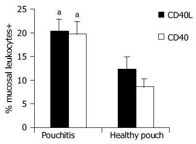
Figure 2 Comparison of CD40L and CD40 leukocytes’ expression between pouchitis and healthy pouch mucosa.
aP < 0.05 vs healthy pouch.
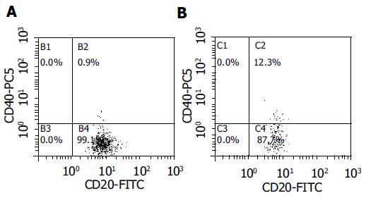
Figure 3 CD40 expression in B cells (CD20+) in controls (A) is homogeneously distributed under the cut-off.
This expression is instead above the cut-off value, in several B cells in pouch patients with spondyloarthropathy (B), as in those with UC.
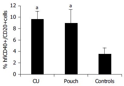
Figure 4 Flow cytometric analysis of CD40+/CD20+ cells in blood.
aP < 0.05 vs controls.
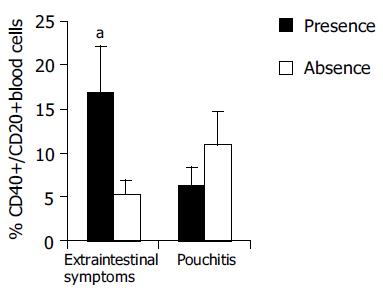
Figure 5 Blood B cells in pouch patients with extraintestinal manifestations, such as spondyloarthropathy or pyoderma gangrenosum, present a higher expression of CD40 with respect to the other pouch patients.
Instead no difference is present during pouchitis. Hfi, high fluorescence intensity. aP < 0.05 vs absence.
- Citation: Polese L, Angriman I, Franchis GD, Cecchetto A, Sturniolo GC, D’Incà R, Scarpa M, Ruffolo C, Norberto L, Frego M, D’Amico DF. Persistence of high CD40 and CD40L expression after restorative proctocolectomy for ulcerative colitis. World J Gastroenterol 2005; 11(34): 5303-5308
- URL: https://www.wjgnet.com/1007-9327/full/v11/i34/5303.htm
- DOI: https://dx.doi.org/10.3748/wjg.v11.i34.5303


