Copyright
©The Author(s) 2005.
World J Gastroenterol. Sep 7, 2005; 11(33): 5229-5231
Published online Sep 7, 2005. doi: 10.3748/wjg.v11.i33.5229
Published online Sep 7, 2005. doi: 10.3748/wjg.v11.i33.5229
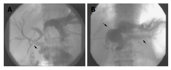
Figure 1 Percutaneous transhepatic cholangiography.
A: A longitudinal stenosis affecting the area of the left bilio-enteric anastomosis is shown. The left bile ducts are markedly dilated; B: Percutaneous transhepatic cholangiography exhibits the extraductal bile collection in the form of a biloma (small arrow) and the dilatation of the left lobe bile ducts (big arrow).
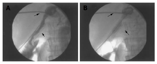
Figure 2 Bile stasis, secondary to stent lumen stenosis (small arrow, A), was managed with external drainage of the biloma (thin arrow, A, B) combined, in the same session, with transhepatic balloon-assisted stent re-dilatation (B).
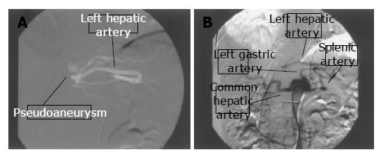
Figure 3 Catheter-directed, selective angiography demonstrates the presence of a pseudoaneurysm arising from the left hepatic artery.
Note that the left hepatic artery originates from the left gastric artery (A and B ).
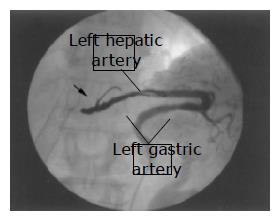
Figure 4 Trans catheter embolization.
Detachment of adequate metallic coils (arrow) resulted in full occlusion of the aneurysmal sac.
- Citation: Siablis D, Papathanassiou ZG, Karnabatidis D, Christeas N, Vagianos C. Hemobilia secondary to hepatic artery pseudoaneurysm: An unusual complication of bile leakage in a patient with a history of a resected IIIb Klatskin tumor. World J Gastroenterol 2005; 11(33): 5229-5231
- URL: https://www.wjgnet.com/1007-9327/full/v11/i33/5229.htm
- DOI: https://dx.doi.org/10.3748/wjg.v11.i33.5229


