Copyright
©The Author(s) 2005.
World J Gastroenterol. Aug 28, 2005; 11(32): 5082-5083
Published online Aug 28, 2005. doi: 10.3748/wjg.v11.i32.5082
Published online Aug 28, 2005. doi: 10.3748/wjg.v11.i32.5082
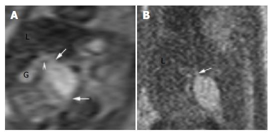
Figure 1 MRI performed at 26 wk’ gestation using HASTE sequence.
A: Coronal T2-weighed image showed the choledochal cyst, its tapered ends (arrows), its connection to the cystic duct (arrowhead), and its relationship to the gall bladder (G) and the liver (L); B: Sagittal T2-weighed image showed the posterio-inferior orientation of the choledochal cyst and its connection to the common hepatic duct (arrow).
- Citation: Wong AMC, Cheung YC, Liu YH, Ng KK, Chan SC, Ng SH. Prenatal diagnosis of choledochal cyst using magnetic resonance imaging: A case report. World J Gastroenterol 2005; 11(32): 5082-5083
- URL: https://www.wjgnet.com/1007-9327/full/v11/i32/5082.htm
- DOI: https://dx.doi.org/10.3748/wjg.v11.i32.5082


