Copyright
©The Author(s) 2005.
World J Gastroenterol. Aug 14, 2005; 11(30): 4667-4673
Published online Aug 14, 2005. doi: 10.3748/wjg.v11.i30.4667
Published online Aug 14, 2005. doi: 10.3748/wjg.v11.i30.4667
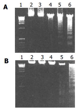
Figure 1 Measurement of apoptosis by DNA fragmentation upon treatment with DCVC/DPPD.
A: SMMC-7721 Hepatocellular carcinoma cells were treated with 0.1 mmol/LDCVC and 0.02 mmol/L DPPD for various time duration, then harvested for DNA fragmentation assay to estimate apoptosis. Lane 1, DNA marker; lane 2, absent DCVC; lanes 3-6 represent tumor cells treated for 1, 2, 4, 6, and 8 h, respectively; B: SMMC-7721 hepatocellular carcinoma cells were treated with 0.02 mmol/L DPPD and various DCVC concentrations for 6 h, then harvested for DNA fragmentation assay to estimate apoptosis. Lane 1, DNA marker; lane 2, absent DCVC; lanes 3–6 represent tumor cells treated for 0.02, 0.05, 0.1, and 0.2 mmol/L, respectively.
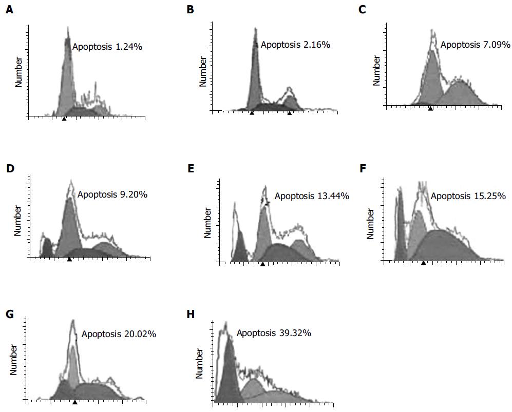
Figure 2 Dose effect on cell apoptosis induced by DCVC co-treated with DPPD.
SMMC-7721 hepatocellular carcinoma cells were treated with 0.02 mmol/L DPPD and various DCVC concentration for 6 h, then harvested for flow cytometry assay to estimate apoptosis. A: Absent both DCVC and DPPD; B: absent DCVC; C: DCVC (0.005 mmol/L); D: DCVC (0.01 mmol/L); E: DCVC (0.02 mmol/L); F: DCVC (0.05 mmol/L); G: DCVC (0.1 mmol/L); H: DCVC (0.2 mmol/L).
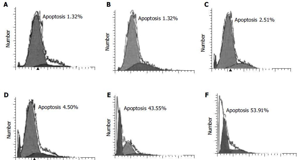
Figure 3 Time effect on cell apoptosis induced by DCVC co-treated with DPPD.
SMMC-7721 hepatocellular carcinoma cells were treated with 0.1 mmol/L DCVC and 0.02 mmol/L DPPD for various time, then harvested for flow cytometry assay to estimate apoptosis. A: absent DCVC, for 8 h; B: for 1 h; C: 2 h; D: 4 h; E: 6 h; F: 8 h.
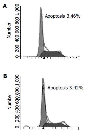
Figure 4 Role of DPPD in apoptosis of SMMC-7721 hepatocellular carcinoma cell.
Cells were cultured in completed medium for 12 h. A: In the absence of DPPD; B: in the presence of 0.02 mmol/L DPPD, then cells were carried out in FMC to estimate apoptosis.
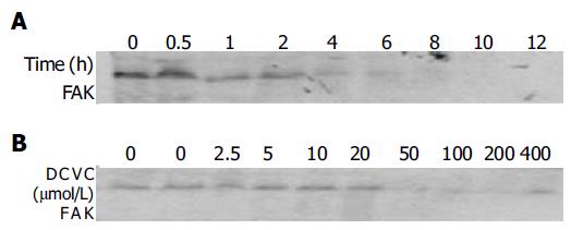
Figure 5 DCVC effects on FAK protein expression.
A: SMMC-7721 hepatocellular carcinoma cells were treated with 0.1 mmol/L DCVC and 0.02 mmol/L DPPD for various time duration; B: SMMC-7721 hepatocellular carcinoma cells were treated with 0.02 mmol/L DPPD and various DCVC concentrations for 6 h, from right the first lane both DCVC and DPPD were absent, the second lane had DPPD but no DCVC. Cells were harvested with 1× SDS loading buffer, followed by Western blot analysis. Equal amounts of cell protein (60 μg) were added to each lane, these studies were repeated thrice with similar results.
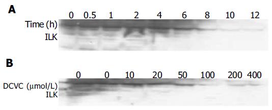
Figure 6 DCVC effects on ILK protein expression.
A: SMMC-7721 hepatocellular carcinoma cells were treated with 0.1 mmol/L DCVC and 0.02 mmol/L DPPD for various time duration; B: SMMC-7721 hepatocellular carcinoma cells were treated with 0.02 mmol/L DPPD and various DCVC concentrations for 6 h, from left the both DCVC and DPPD were absent, the second lane had DPPD but no DCVC. Cells were harvested with 1× SDS loading buffer, followed by Western blot analysis. Equal amounts of cell protein (60 μg) were added to each lane, these studies were repeated thrice with similar results.
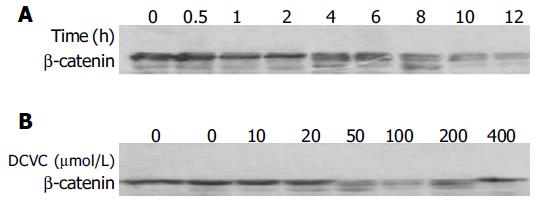
Figure 7 DCVC effects on β-catenin protein expression.
A: SMMC-7721 hepatocellular carcinoma cells were treated with 0.1 mmol/L DCVC and 0.02 mmol/L DPPD for various time duration; B: SMMC-7721 hepatocellular carcinoma cells were treated with 0.02 mmol/L DPPD and various DCVC concentrations for 6 h, from left the first lane both DCVC and DPPD were absent, the second lane had DPPD but no DCVC. Cells were harvested with 1× SDS loading buffer, followed by Western blot analysis. Equal amounts of cell protein (60 μg) were added to each lane, these studies were repeated thrice with similar results.
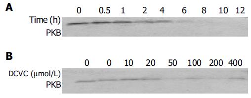
Figure 8 DCVC effects on PKB protein expression.
A: SMMC-7721 hepatocellular carcinoma cells were treated with 0.1 mmol/L DCVC and 0.02 mmol/L DPPD for various time duration; B: SMMC-7721 hepatocellular carcinoma cells were treated with 0.02 mmol/L DPPD and various DCVC concentration for 6 h, from left both DCVC and DPPD were absent in the first lane, the second lane had DPPD but without DCVC. Cells were harvested with 1× SDS loading buffer, followed by Western blot analysis. Equal amounts of cell protein (60 μg) were added to each lane, these studies were repeated thrice with similar results.
- Citation: Su JM, Wang LY, Liang YL, Zha XL. Role of cell adhesion signal molecules in hepatocellular carcinoma cell apoptosis. World J Gastroenterol 2005; 11(30): 4667-4673
- URL: https://www.wjgnet.com/1007-9327/full/v11/i30/4667.htm
- DOI: https://dx.doi.org/10.3748/wjg.v11.i30.4667


